September 4, 2024
Optimal fixation, processing, and embedding of Formalin-Fixed, Paraffin-Embedded (FFPE) tissue are crucial steps in histological examination. These initial stages ensure high-quality staining, imaging, and quantitative analysis of tissue morphology and biomarker localization. This guide provides an overview of common fixation, processing, and embedding methods for FFPE rodent tissue.
Proper tissue fixation preserves the tissue architecture, including cells, cellular components (cytoplasm, nuclei, organelles), extracellular material, and molecular components (proteins, DNA, mRNA). Various tissue fixatives are available, each with specific advantages and disadvantages depending on the application. Timing is also key. The cold ischemia time, time from removal of tissue until placement in fixative, should not exceed 1 hour.
10% NBF is the most widely used fixative in routine diagnostic pathology due to its ability to preserve tissue effectively. Key factors for optimal results include:
For tissues like eyes, lungs, intestines, and bones that may require different fixatives or special processing, contact our expert team for guidance on the appropriate tissue fixation techniques.
Fixed tissues are typically processed using automated tissue processors and embedded in paraffin wax. This process involves immersing samples in a series of solutions, including alcohols, xylene, and paraffin wax. Customization of each step—time, temperature, and pressure/vacuum (P/V)—ensures optimal solution penetration.
Our histologists have developed specific tissue processing schedules tailored to different tissue types, ensuring high-quality outcomes. Understanding the steps of tissue processing is essential for maintaining the integrity of FFPE samples.
High-quality embedding requires consistent orientation of all samples of the same tissue type. While samples can be embedded in various orientations upon request, we typically follow the Registry of Industrial Toxicology Animal (RITA) and North American Control Animal Database (NACAD) Guidelines for rodent tissue embedding.
If your study requires a different tissue embedding orientation, please specify in advance, and we will accommodate your needs.
Optimal fixation, processing, and embedding are critical steps for examining tissue morphology and biomarker localization in FFPE tissue samples. Our expert team can assist you with all aspects of formalin-fixed paraffin-embedded tissue preparation. Contact us if you have any questions, our team is here to ensure your histology workflow is optimized for the best results.
September 4, 2024
Optimal fixation, processing, and embedding of Formalin-Fixed, Paraffin-Embedded (FFPE) tissue are crucial steps in histological examination. These initial stages ensure high-quality staining, imaging, and quantitative analysis of tissue morphology and biomarker localization. This guide provides an overview of common fixation, processing, and embedding methods for FFPE rodent tissue.
Proper tissue fixation preserves the tissue architecture, including cells, cellular components (cytoplasm, nuclei, organelles), extracellular material, and molecular components (proteins, DNA, mRNA). Various tissue fixatives are available, each with specific advantages and disadvantages depending on the application. Timing is also key. The cold ischemia time, time from removal of tissue until placement in fixative, should not exceed 1 hour.
10% NBF is the most widely used fixative in routine diagnostic pathology due to its ability to preserve tissue effectively. Key factors for optimal results include:
For tissues like eyes, lungs, intestines, and bones that may require different fixatives or special processing, contact our expert team for guidance on the appropriate tissue fixation techniques.
Fixed tissues are typically processed using automated tissue processors and embedded in paraffin wax. This process involves immersing samples in a series of solutions, including alcohols, xylene, and paraffin wax. Customization of each step—time, temperature, and pressure/vacuum (P/V)—ensures optimal solution penetration.
Our histologists have developed specific tissue processing schedules tailored to different tissue types, ensuring high-quality outcomes. Understanding the steps of tissue processing is essential for maintaining the integrity of FFPE samples.
High-quality embedding requires consistent orientation of all samples of the same tissue type. While samples can be embedded in various orientations upon request, we typically follow the Registry of Industrial Toxicology Animal (RITA) and North American Control Animal Database (NACAD) Guidelines for rodent tissue embedding.
If your study requires a different tissue embedding orientation, please specify in advance, and we will accommodate your needs.
Optimal fixation, processing, and embedding are critical steps for examining tissue morphology and biomarker localization in FFPE tissue samples. Our expert team can assist you with all aspects of formalin-fixed paraffin-embedded tissue preparation. Contact us if you have any questions, our team is here to ensure your histology workflow is optimized for the best results.
May 28, 2024
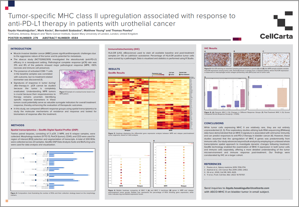
CellCarta’s poster, presented at ASCO 2024, discusses the study on tumor-specific MHC class II upregulation in patients with urothelial cancer undergoing anti-PD-L1 therapy.
The study, part of the abacus trial (NCT02662309), revealed that MHC class II expression in tumor cells, although rare, is associated with a positive response to atezolizumab. Utilizing spatial transcriptomics and immunohistochemistry (IHC), the research highlighted significant differences in MHC II expression between patients with pathological complete response (pCR) and those with major pathological response (MPR) or relapse.
The findings suggest that MHC II expression could serve as a biomarker for predicting therapeutic outcomes in bladder cancer treatment.
View the full poster:
May 28, 2024
CellCarta’s poster, presented at ASCO 2024, discusses the study on tumor-specific MHC class II upregulation in patients with urothelial cancer undergoing anti-PD-L1 therapy.
The study, part of the abacus trial (NCT02662309), revealed that MHC class II expression in tumor cells, although rare, is associated with a positive response to atezolizumab. Utilizing spatial transcriptomics and immunohistochemistry (IHC), the research highlighted significant differences in MHC II expression between patients with pathological complete response (pCR) and those with major pathological response (MPR) or relapse.
The findings suggest that MHC II expression could serve as a biomarker for predicting therapeutic outcomes in bladder cancer treatment.
View the full poster: