Optimizing your in situ hybridization for success

August 30, 2024

What is In Situ Hybridization (ISH)?

in situ hybridization (ISH) is a technique that enables the localization of specific DNA or RNA sequences within tissue sections and cytology samples using labeled complementary strands. The target gene can be visualized through brightfield or fluorescence microscopy. Compared to immunohistochemistry (IHC), which broadly localizes proteins within tissues, ISH offers greater sensitivity, allowing researchers to understand the organization and function of genes in tissue sections. ISH not only identifies cells positive for a specific DNA/RNA sequence but also quantifies the level of positivity within individual cells.

How can you benefit from combining ISH and IHC assays

ISH is a powerful tool in both preclinical research and clinical trials, offering critical insights. It can be combined with IHC assays to provide unique perspectives on cellular and molecular mechanisms within tissues.

Key applications include:

  • Relationship Between Targets: Dual assays on unique but related targets reveal their interrelationships.
  • Protein Localization: Identify the location of a target protein within tissue and the originating cells.
  • Gene Regulation Insight: Combining ISH and IHC on the same target provides insights into gene regulation mechanisms.
  • Tumor Region Analysis: Use IHC cytokeratin markers with ISH assays to highlight gene regulation in tumor regions.
  • Protein Degradation Insights: Dual assays offer valuable insights into protein degradation processes.

Maximizing your in situ hybridization assays

Successful ISH assays depend on meticulous sample preparation to minimize RNA degradation in tissue.

  • Tissue Fixation: Fix tissues promptly in 10% Neutral Buffered Formalin (NBF) at 20x the tissue sample volume, then transfer to 70% ethanol (EtOH).
  • RNA/DNA Preservation: Cut fresh sections under RNase/DNase-free conditions onto charged slides for better adhesion, and store at a constant 4°C or at room temperature.
  •  Baking conditions: Bake slides for 30-120 min at 60°C for better adhesion.

Another crucial aspect for a successful ISH assay is optimization. Each unique combination of species, tissue type, and gene target must be optimized to hone in on the ideal conditions for detecting and visualizing the target. Pre-existing IHC data can guide the optimization process.

Additionally, digital whole slide imaging settings must be tailored to each sample set, accounting for tissue type differences, stain intensity, and autofluorescence.

Consistency in applying the ISH protocol across samples is key. Automated technologies, like the Leica BondRX and Ventana Discovery Ultra, ensure precise and consistent application, minimizing human error.

Leveraging AI-Powered Data for your ISH Assays

The integration of AI-powered algorithms with digital whole slide ISH images enhances the analysis, generating data and insights beyond human capability. However, several key steps ensure successful ISH assays.

Our AI-powered algorithms ensure optimal nuclear segmentation for the most reliable results.

Rest assured that our in-house team of pathologists work in proximity with our imaging specialists to ensure you get optimized data from your ISH assays, that signal is neither undercounted nor overestimated.

By leveraging cutting-edge ISH technologies, CellCarta ensures comprehensive and high-quality results for your research and diagnostic needs.

Contact us to get your project started.


Our expert:

author photo

Sofie Van Rossom (PhD) is a scientific matter expert for ISH technologies within CellCarta. With a background is Biochemistry & Biotechnology, and many years of experience in the development and validation of new (F)ISH assays  to identify DNA and RNA biomarkers of clinical utility, Sofie uses her expertise to guide customers in the validation of their (F)ISH assays.

Optimizing your in situ hybridization for success

August 30, 2024

What is In Situ Hybridization (ISH)?

in situ hybridization (ISH) is a technique that enables the localization of specific DNA or RNA sequences within tissue sections and cytology samples using labeled complementary strands. The target gene can be visualized through brightfield or fluorescence microscopy. Compared to immunohistochemistry (IHC), which broadly localizes proteins within tissues, ISH offers greater sensitivity, allowing researchers to understand the organization and function of genes in tissue sections. ISH not only identifies cells positive for a specific DNA/RNA sequence but also quantifies the level of positivity within individual cells.

How can you benefit from combining ISH and IHC assays

ISH is a powerful tool in both preclinical research and clinical trials, offering critical insights. It can be combined with IHC assays to provide unique perspectives on cellular and molecular mechanisms within tissues.

Key applications include:

  • Relationship Between Targets: Dual assays on unique but related targets reveal their interrelationships.
  • Protein Localization: Identify the location of a target protein within tissue and the originating cells.
  • Gene Regulation Insight: Combining ISH and IHC on the same target provides insights into gene regulation mechanisms.
  • Tumor Region Analysis: Use IHC cytokeratin markers with ISH assays to highlight gene regulation in tumor regions.
  • Protein Degradation Insights: Dual assays offer valuable insights into protein degradation processes.

Maximizing your in situ hybridization assays

Successful ISH assays depend on meticulous sample preparation to minimize RNA degradation in tissue.

  • Tissue Fixation: Fix tissues promptly in 10% Neutral Buffered Formalin (NBF) at 20x the tissue sample volume, then transfer to 70% ethanol (EtOH).
  • RNA/DNA Preservation: Cut fresh sections under RNase/DNase-free conditions onto charged slides for better adhesion, and store at a constant 4°C or at room temperature.
  •  Baking conditions: Bake slides for 30-120 min at 60°C for better adhesion.

Another crucial aspect for a successful ISH assay is optimization. Each unique combination of species, tissue type, and gene target must be optimized to hone in on the ideal conditions for detecting and visualizing the target. Pre-existing IHC data can guide the optimization process.

Additionally, digital whole slide imaging settings must be tailored to each sample set, accounting for tissue type differences, stain intensity, and autofluorescence.

Consistency in applying the ISH protocol across samples is key. Automated technologies, like the Leica BondRX and Ventana Discovery Ultra, ensure precise and consistent application, minimizing human error.

Leveraging AI-Powered Data for your ISH Assays

The integration of AI-powered algorithms with digital whole slide ISH images enhances the analysis, generating data and insights beyond human capability. However, several key steps ensure successful ISH assays.

Our AI-powered algorithms ensure optimal nuclear segmentation for the most reliable results.

Rest assured that our in-house team of pathologists work in proximity with our imaging specialists to ensure you get optimized data from your ISH assays, that signal is neither undercounted nor overestimated.

By leveraging cutting-edge ISH technologies, CellCarta ensures comprehensive and high-quality results for your research and diagnostic needs.

Contact us to get your project started.


Our expert:

author photo

Sofie Van Rossom (PhD) is a scientific matter expert for ISH technologies within CellCarta. With a background is Biochemistry & Biotechnology, and many years of experience in the development and validation of new (F)ISH assays  to identify DNA and RNA biomarkers of clinical utility, Sofie uses her expertise to guide customers in the validation of their (F)ISH assays.
Cell Doublets in Flow Cytometry Data | Benefits & Challenges CellCarta

August 15, 2024

doublets in flow cytometry

Flow cytometry (including cell sorting) provides unprecedented levels of detail about blood samples for both research and clinical applications, allowing for instance the unraveling of phenotypic heterogeneity of human immune cells.

However, research from the Burel group , La Jolla Institute for Immunology, highlighted that blood-derived cell doublets can be present in single-cell data derived from flow cytometry analyses.

Doublet exclusion methods, commonly based on Forward Scatter (FSC)/Side Scatter (SSC) Area/Width/Height measurements, can partly remove cell doublets, but some complexes remain undetected, leading to data misinterpretation.

While cell doublets often need removing, interestingly, Burel reported that some doublets form naturally — which could provide greater disease understanding.

Here we look at what cell doublets are, their risk and promise, and how analysts can ensure accurate results.

Seeing Double: Identifying Cell Doublets in Flow Cytometry

While dual lineage co-expressing cell populations (also called dual expressor cells) exist in human peripheral blood samples analysed by flow cytometry, Burel’s team recently unveiled through imaging that some of these events are actually cell-cell complexes.

In the live singlet gate of blood-derived samples, T cells can still be bound to other immune cells, such as antigen-presenting cells (APC), de facto forming a cell doublet. But without deeper analysis, it can be easy to analyze cell doublets as single cells.

In cell sorting applications, the cell doublets can subsequently generate atypical gene signatures of mixed cell lineage, meaning that data misinterpretation can occur, including:

  • Specific cell population percentage deflation,
  • Novel immune cell type misidentification, and
  • Incorrect functional marker expression attribution.

Distinguishing cell-cell complexes for more accurate Flow Cytometry results

Experimental and data analysis strategies can distinguish between singlets and cell doublets for more accurate data.

To detect the presence of cell-cell complexes within potential dual-expressing cells, Burel’s team used two different approaches:

  • Cell sorting followed by direct microscopy imaging to confirm the expression of markers on distinct cells
  • Imaging flow cytometry to generate metrics from pictures of the detected events, enabling doublet exclusion using brightfield area and aspect ratio parameter calculation.

Notably, Burel’s team found that cell-cell complexes have molecular signatures that clearly distinguish them from singlets at both protein and mRNA levels, providing analysts with a way to avoid data misinterpretation.

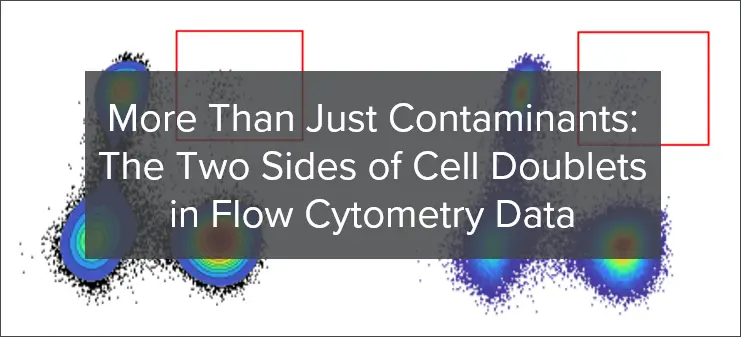
When looking at cell–cell complex signatures with SSC/FSC and SSC/CD45, the team discovered that the complexes had higher levels of CD45, SSC, and FSC expression compared to single cells.

Importantly, cell doublets aren’t always just contaminants. The team found that T cell-APC complexes that form in vivo as a natural immunological process can be detected in the singlet gate of ex vivo human blood samples analyzed by flow cytometry, in quantities that vary over time depending on the disease state.

As such, it is therefore believed that they could provide deeper insights into research and diagnosis.

Integration of Techniques from Doublet Discrimination and Exclusion: Techniques and Detailed Process

Practical Applications of Doublet Discrimination in Flow Cytometry Analyses

Proper doublet discrimination ensures increased data accuracy and enhanced data quality. Therefore, proper singlet gating strategies in flow cytometry analyses are essential for ensuring that only single-cell events are analyzed, leading to more reliable and reproducible results.

There are essentially two main techniques for identifying and gating singlet events in flow cytometry data:

  •  Pulse Processing: incorporating Area, Height and Width bi-variate plots for FSC and SSC parameters in the hierarchical gating strategy can help identify and exclude doublets based in their distinct scatter profiles.
  •  Autofluorescence extraction: in full-spectrum cytometry, a technology that captures the entire fluorescence spectrum emitted by each fluorochrome, the autofluorescence of the sample (natural emission of light by biological structures) can be extracted as well and used to discriminate doublets. This is particularly useful for analyzing tissues or cells with high levels of autofluorescence, such as lung tissues or certain cell types in immunology and oncology research.

Singlet vs. Doublet Flow Cytometry Analysis

Understanding the differences between singlets and doublets is crucial for precise flow cytometry analysis. The following table highlights key distinctions:

Feature Singlet Flow Cytometry Doublet Flow Cytometry
Cell Type Single cells Doublet cells
Identification Method Linear relationship in FSC and SSC pulse dimensions (Area, Height, Width) Deviations from linear relationship
Data Accuracy High Can lead to data misinterpretation
Analysis Focus Individual cell analysis Potentially confounded by cell aggregates
Impact on Results Accurate cell population quantification Potentially confounded by cell aggregates

Leverage expertise for maximum data confidence in Flow Cytometry

Addressing the issue of cell doublets in flow cytometry is essential for maintaining data accuracy and quality.

By employing effective doublet discrimination and singlet gating techniques, researchers can ensure that their flow cytometry data reflects true single-cell events. This leads to more reliable and reproducible results, ultimately advancing our understanding of complex biological systems.

Our analytical team has the expertise to tailor gating strategies based on the presence of cell-cell complex doublets. CellCarta offers a broad range of advanced services using robust techniques to get greater insights from your studies. Contact the team to learn more.


About the author:

author photo

Roxanne Collin is a Research Scientist within the Development group of the Data Analysis Unit at CellCarta. She holds a PhD in immunology with a focus on the immunogenetics of rare cell populations. At CellCarta, she is participating in the optimisation of flow cytometry panels and gating strategies for a broad variety of immune cell types.

Cell Doublets in Flow Cytometry Data | Benefits & Challenges CellCarta

August 15, 2024

doublets in flow cytometry

Flow cytometry (including cell sorting) provides unprecedented levels of detail about blood samples for both research and clinical applications, allowing for instance the unraveling of phenotypic heterogeneity of human immune cells.

However, research from the Burel group , La Jolla Institute for Immunology, highlighted that blood-derived cell doublets can be present in single-cell data derived from flow cytometry analyses.

Doublet exclusion methods, commonly based on Forward Scatter (FSC)/Side Scatter (SSC) Area/Width/Height measurements, can partly remove cell doublets, but some complexes remain undetected, leading to data misinterpretation.

While cell doublets often need removing, interestingly, Burel reported that some doublets form naturally — which could provide greater disease understanding.

Here we look at what cell doublets are, their risk and promise, and how analysts can ensure accurate results.

Seeing Double: Identifying Cell Doublets in Flow Cytometry

While dual lineage co-expressing cell populations (also called dual expressor cells) exist in human peripheral blood samples analysed by flow cytometry, Burel’s team recently unveiled through imaging that some of these events are actually cell-cell complexes.

In the live singlet gate of blood-derived samples, T cells can still be bound to other immune cells, such as antigen-presenting cells (APC), de facto forming a cell doublet. But without deeper analysis, it can be easy to analyze cell doublets as single cells.

In cell sorting applications, the cell doublets can subsequently generate atypical gene signatures of mixed cell lineage, meaning that data misinterpretation can occur, including:

  • Specific cell population percentage deflation,
  • Novel immune cell type misidentification, and
  • Incorrect functional marker expression attribution.

Distinguishing cell-cell complexes for more accurate Flow Cytometry results

Experimental and data analysis strategies can distinguish between singlets and cell doublets for more accurate data.

To detect the presence of cell-cell complexes within potential dual-expressing cells, Burel’s team used two different approaches:

  • Cell sorting followed by direct microscopy imaging to confirm the expression of markers on distinct cells
  • Imaging flow cytometry to generate metrics from pictures of the detected events, enabling doublet exclusion using brightfield area and aspect ratio parameter calculation.

Notably, Burel’s team found that cell-cell complexes have molecular signatures that clearly distinguish them from singlets at both protein and mRNA levels, providing analysts with a way to avoid data misinterpretation.

When looking at cell–cell complex signatures with SSC/FSC and SSC/CD45, the team discovered that the complexes had higher levels of CD45, SSC, and FSC expression compared to single cells.

Importantly, cell doublets aren’t always just contaminants. The team found that T cell-APC complexes that form in vivo as a natural immunological process can be detected in the singlet gate of ex vivo human blood samples analyzed by flow cytometry, in quantities that vary over time depending on the disease state.

As such, it is therefore believed that they could provide deeper insights into research and diagnosis.

Integration of Techniques from Doublet Discrimination and Exclusion: Techniques and Detailed Process

Practical Applications of Doublet Discrimination in Flow Cytometry Analyses

Proper doublet discrimination ensures increased data accuracy and enhanced data quality. Therefore, proper singlet gating strategies in flow cytometry analyses are essential for ensuring that only single-cell events are analyzed, leading to more reliable and reproducible results.

There are essentially two main techniques for identifying and gating singlet events in flow cytometry data:

  •  Pulse Processing: incorporating Area, Height and Width bi-variate plots for FSC and SSC parameters in the hierarchical gating strategy can help identify and exclude doublets based in their distinct scatter profiles.
  •  Autofluorescence extraction: in full-spectrum cytometry, a technology that captures the entire fluorescence spectrum emitted by each fluorochrome, the autofluorescence of the sample (natural emission of light by biological structures) can be extracted as well and used to discriminate doublets. This is particularly useful for analyzing tissues or cells with high levels of autofluorescence, such as lung tissues or certain cell types in immunology and oncology research.

Singlet vs. Doublet Flow Cytometry Analysis

Understanding the differences between singlets and doublets is crucial for precise flow cytometry analysis. The following table highlights key distinctions:

Feature Singlet Flow Cytometry Doublet Flow Cytometry
Cell Type Single cells Doublet cells
Identification Method Linear relationship in FSC and SSC pulse dimensions (Area, Height, Width) Deviations from linear relationship
Data Accuracy High Can lead to data misinterpretation
Analysis Focus Individual cell analysis Potentially confounded by cell aggregates
Impact on Results Accurate cell population quantification Potentially confounded by cell aggregates

Leverage expertise for maximum data confidence in Flow Cytometry

Addressing the issue of cell doublets in flow cytometry is essential for maintaining data accuracy and quality.

By employing effective doublet discrimination and singlet gating techniques, researchers can ensure that their flow cytometry data reflects true single-cell events. This leads to more reliable and reproducible results, ultimately advancing our understanding of complex biological systems.

Our analytical team has the expertise to tailor gating strategies based on the presence of cell-cell complex doublets. CellCarta offers a broad range of advanced services using robust techniques to get greater insights from your studies. Contact the team to learn more.


About the author:

author photo

Roxanne Collin is a Research Scientist within the Development group of the Data Analysis Unit at CellCarta. She holds a PhD in immunology with a focus on the immunogenetics of rare cell populations. At CellCarta, she is participating in the optimisation of flow cytometry panels and gating strategies for a broad variety of immune cell types.

Why drug development needs spatial biology

August 13, 2024

Why drug development needs spatial biology

Understanding how drug candidates interact within diverse cellular environments and how these interactions translate to patient outcomes can be challenging. Moreover, heterogeneity within seemingly uniform diseases can lead to varied treatment responses, complicating the path to truly personalized therapies. As the field grapples with these challenges, new methodologies are needed to unravel the intricacies of disease and drug response. To this end, attention is increasingly turning to spatial biology.

What is spatial biology?

Spatial biology is the study of cells within their native environments. Unlike traditional bulk approaches, spatial biology preserves the spatial context, allowing scientists to study how cells and molecules are distributed and interact within complex tissues and the architectural changes associated with various biological processes and diseases. By leveraging these insights, spatial biology enhances the understanding of drug effects at the cellular level, aiding the development of targeted therapies.

While spatial biology is not a new concept recent advances in multiplexing have transformed the field. These innovations enable the simultaneous measurement of multiple markers, and with enhanced computational capabilities, we can more effectively analyze the relationships between them. This deeper understanding of complex biological paradigms has elevated spatial biology into a powerful tool for drug development.

Why drug developers are turning to spatial biology

Drug developers are turning to spatial biology because it offers a deeper understanding of cellular context, leading to more accurate drug targeting, better predictions of drug efficacy, and insights into disease mechanisms.

As drug development is complex and costly, these comprehensive tools are essential for improving the efficiency of therapy development. Integrating spatial biology into drug development workflows offers several benefits:

  • Aids understanding of tissue microenvironments: Spatial biology can reveal the complexities of the tissue microenvironment, leading to the identification of new therapeutic targets and treatments.
  • Addresses disease heterogeneity: Spatial biology enables the examination of the cellular heterogeneity that traditional bulk methods might miss. This helps unravel how cells organize and contribute to disease progression and treatment response.
  • Enables biomarker identification: Spatial biology aids biomarker identification by revealing cells’ exact location and interactions within their native tissue environments, giving a detailed view of cellular behavior and disease mechanisms.
  • Enhances drug distribution and efficacy studies: Spatial biology provides insights into the pharmacodynamics and pharmacokinetics of new compounds.
  •  Elucidates mechanism of action: Spatial biology provides a detailed understanding of how cells interact and respond to treatments, thereby clarifying drugs’ mechanisms of action.
  •  Improves immune response profiling: Spatial biology enhances the ability to profile immune responses by accurately mapping the infiltration status and immune cell states within their native tissue context.
  • Builds understanding of drug resistance: Spatial biology helps to unravel the mechanisms behind drug resistance by examining how cellular and tissue environments influence the effectiveness of therapies.

A discovery toolbox to accelerate drug development

Spatial biology has emerged as a groundbreaking discovery toolbox, significantly accelerating drug development by offering detailed insights into tissue microenvironments and molecular interactions. Cutting-edge platforms like Visium and Visium HD from 10X Genomics, GeoMX Digital Spatial Profiler from NanoString, and COMET from Lunaphore are at the forefront of this revolution. These platforms allow researchers to simultaneously capture high-plex RNA and protein information from a single tissue section, enabling a comprehensive understanding of cellular contexts and interactions.

Visium HD delivers high-resolution spatially resolved gene expression profiles, while GeoMX offers high-plex protein and RNA analysis powered by precise segmentation capabilities. COMET complements these with its sub-cellular resolution imaging capabilities and allows for the detailed exploration of tissue architecture and biomolecular interactions.

These tools are crucial for early clinical research as they rapidly generate valuable insights into disease mechanisms, drug efficacy, and biomarker validation.

Accelerating your drug development with spatial biology

By facilitating the exploration of complex tissue environments and enabling a deeper understanding of cellular heterogeneity, spatial biology tools streamline the path from preclinical studies to clinical application. This accelerates the identification of promising therapeutic candidates and optimizes their development, ensuring that drug developers can address key challenges more efficiently. With the path to clinical implementation becoming increasingly accessible, these advanced spatial biology platforms are poised to play a pivotal role in shaping the future of personalized medicine and therapeutic innovation.

To learn more about spatial biology and how it could help support and accelerate your therapeutic development, visit: spatial biology

About the author

author photo

İlayda Hasakioğulları is a Senior R&D Product Manager at CellCarta. Ilayda is passionate about precision medicine and specializes in spatial biology technologies. She continually scans the landscape to implement cutting-edge solutions at CellCarta and assists scientists in selecting the best tools for their research needs with the ultimate goal of benefiting patients.

Why drug development needs spatial biology

August 13, 2024

Why drug development needs spatial biology

Understanding how drug candidates interact within diverse cellular environments and how these interactions translate to patient outcomes can be challenging. Moreover, heterogeneity within seemingly uniform diseases can lead to varied treatment responses, complicating the path to truly personalized therapies. As the field grapples with these challenges, new methodologies are needed to unravel the intricacies of disease and drug response. To this end, attention is increasingly turning to spatial biology.

What is spatial biology?

Spatial biology is the study of cells within their native environments. Unlike traditional bulk approaches, spatial biology preserves the spatial context, allowing scientists to study how cells and molecules are distributed and interact within complex tissues and the architectural changes associated with various biological processes and diseases. By leveraging these insights, spatial biology enhances the understanding of drug effects at the cellular level, aiding the development of targeted therapies.

While spatial biology is not a new concept recent advances in multiplexing have transformed the field. These innovations enable the simultaneous measurement of multiple markers, and with enhanced computational capabilities, we can more effectively analyze the relationships between them. This deeper understanding of complex biological paradigms has elevated spatial biology into a powerful tool for drug development.

Why drug developers are turning to spatial biology

Drug developers are turning to spatial biology because it offers a deeper understanding of cellular context, leading to more accurate drug targeting, better predictions of drug efficacy, and insights into disease mechanisms.

As drug development is complex and costly, these comprehensive tools are essential for improving the efficiency of therapy development. Integrating spatial biology into drug development workflows offers several benefits:

  • Aids understanding of tissue microenvironments: Spatial biology can reveal the complexities of the tissue microenvironment, leading to the identification of new therapeutic targets and treatments.
  • Addresses disease heterogeneity: Spatial biology enables the examination of the cellular heterogeneity that traditional bulk methods might miss. This helps unravel how cells organize and contribute to disease progression and treatment response.
  • Enables biomarker identification: Spatial biology aids biomarker identification by revealing cells’ exact location and interactions within their native tissue environments, giving a detailed view of cellular behavior and disease mechanisms.
  • Enhances drug distribution and efficacy studies: Spatial biology provides insights into the pharmacodynamics and pharmacokinetics of new compounds.
  •  Elucidates mechanism of action: Spatial biology provides a detailed understanding of how cells interact and respond to treatments, thereby clarifying drugs’ mechanisms of action.
  •  Improves immune response profiling: Spatial biology enhances the ability to profile immune responses by accurately mapping the infiltration status and immune cell states within their native tissue context.
  • Builds understanding of drug resistance: Spatial biology helps to unravel the mechanisms behind drug resistance by examining how cellular and tissue environments influence the effectiveness of therapies.

A discovery toolbox to accelerate drug development

Spatial biology has emerged as a groundbreaking discovery toolbox, significantly accelerating drug development by offering detailed insights into tissue microenvironments and molecular interactions. Cutting-edge platforms like Visium and Visium HD from 10X Genomics, GeoMX Digital Spatial Profiler from NanoString, and COMET from Lunaphore are at the forefront of this revolution. These platforms allow researchers to simultaneously capture high-plex RNA and protein information from a single tissue section, enabling a comprehensive understanding of cellular contexts and interactions.

Visium HD delivers high-resolution spatially resolved gene expression profiles, while GeoMX offers high-plex protein and RNA analysis powered by precise segmentation capabilities. COMET complements these with its sub-cellular resolution imaging capabilities and allows for the detailed exploration of tissue architecture and biomolecular interactions.

These tools are crucial for early clinical research as they rapidly generate valuable insights into disease mechanisms, drug efficacy, and biomarker validation.

Accelerating your drug development with spatial biology

By facilitating the exploration of complex tissue environments and enabling a deeper understanding of cellular heterogeneity, spatial biology tools streamline the path from preclinical studies to clinical application. This accelerates the identification of promising therapeutic candidates and optimizes their development, ensuring that drug developers can address key challenges more efficiently. With the path to clinical implementation becoming increasingly accessible, these advanced spatial biology platforms are poised to play a pivotal role in shaping the future of personalized medicine and therapeutic innovation.

To learn more about spatial biology and how it could help support and accelerate your therapeutic development, visit: spatial biology

About the author

author photo

İlayda Hasakioğulları is a Senior R&D Product Manager at CellCarta. Ilayda is passionate about precision medicine and specializes in spatial biology technologies. She continually scans the landscape to implement cutting-edge solutions at CellCarta and assists scientists in selecting the best tools for their research needs with the ultimate goal of benefiting patients.

The role of cytokine profiling in a changing cell therapy testing landscape

August 6, 2024

The cell therapy landscape is evolving at unprecedented pace, with an increasingly complex and diversified catalogue of candidates now reaching the clinic, and urgency to bring new therapies to the market.

Together, these trends are putting an increasing burden on clinical testing programs.

In our recent Cell Therapy Trends Report, our experts discuss in more detail how clinical testing strategies are accommodating these rapid development strategies, as well as what that could mean for key clinical testing areas.

One of these key testing areas — the focus of this blog — is cytokine profiling.

Why is cytokine profiling so important for cell therapy clinical testing?

Throughout cell therapy development, cytokine profiling assays provide a report on various pharmacodynamic aspects of the therapeutic agent, including proliferation and activation of CAR T cells, as well as broader activation of endogenous immune components. Further expansion of CAR T populations can also lead to increased levels of cytokines in peripheral blood as hallmarks of a broader systemic inflammatory response. Unfortunately, this can lead to adverse events in various organ systems including the central nervous system (CNS).

Today, several immunoassay platforms are used for cytokine profiling, each differing in detection signal and advantages for different testing scales and hypotheses. The most common platforms are:

  • Meso Scale Discovery® (MSD): MSD assays use electrochemiluminescent labels and enable ultra-sensitive detection
  •  Olink® PEA Technology: this technology combines an antibody-based immunoassay with polymerase chain reaction (PCR) to provide high-throughput multi-plex analysis
  • Ella™: a next-generation, automated benchtop ELISA multiplexing system that delivers high-speed precision with minimal user input

So, what insights can these assays give to developers, exactly?

When deployed in early clinical phases, cytokine profiling assays can provide crucial insight into the temporal dynamics of immune cell activation and adverse effects, such as cytokine release syndrome (CRS) and immune effector cell-associated neurotoxicity syndrome (ICANS). Combined with other assays (such as cell enumeration, cell phenotyping, and VCN determination), cytokine profiling can also support decision making on optimal dosing levels, reducing the reliance on the maximum tolerable dose.

The future of cytokine profiling: emerging roles and applications

As cell therapies diversify in complexity, target, and agent, the roles and applications of cytokine profiling in cell therapy testing are set to change in several ways.

Assessing novel secondary mechanisms

For every element added to a cell construct, monitoring of a new biomarker or signature is needed to show that the addition has the intended effect in the body. Cytokine profiling is likely to help here, potentially playing a key role in evaluating the secondary mechanisms of next-generation construct designs such as CAR T cell armoring.

Understanding the impact of patient immune status

Another emerging application of cytokine profiling is understanding baseline cytokine signatures that correlate with clinically relevant parameters. With these signatures, researchers can better understand how a patient’s immune status influences the quality and action of cell therapy. Naturally, such knowledge could have far-reaching impacts. For example, insight into how patient health affects therapy efficacy will be paramount for more confidently evaluating therapies approved as earlier-line treatments — treatments that operate in the context of a less depleted immune system.

Staying abreast of trends in cell therapy clinical testing

Keen to find out more about the current and anticipated future applications of cytokine profiling and other key testing areas? Our experts discuss all this and more in our Cell Therapy Trends Report.

Download the report for free today.

Alternatively, contact our team to speak to an expert about your cell therapy needs.

About the author: 

author photo

Nick Dupuis is a Senior Director of Scientific Business Development at CellCarta and has a background in in proteomics and biophysical measurement. He has held various roles in industry including as a scientist in LDT/IVD product and applications development, and business development for emerging analytical platforms. At CellCarta he leads commercialization initiatives touching on multiomic applications for biomarkers in cell therapy and pathways to support companion diagnostic development.

The role of cytokine profiling in a changing cell therapy testing landscape

August 6, 2024

The cell therapy landscape is evolving at unprecedented pace, with an increasingly complex and diversified catalogue of candidates now reaching the clinic, and urgency to bring new therapies to the market.

Together, these trends are putting an increasing burden on clinical testing programs.

In our recent Cell Therapy Trends Report, our experts discuss in more detail how clinical testing strategies are accommodating these rapid development strategies, as well as what that could mean for key clinical testing areas.

One of these key testing areas — the focus of this blog — is cytokine profiling.

Why is cytokine profiling so important for cell therapy clinical testing?

Throughout cell therapy development, cytokine profiling assays provide a report on various pharmacodynamic aspects of the therapeutic agent, including proliferation and activation of CAR T cells, as well as broader activation of endogenous immune components. Further expansion of CAR T populations can also lead to increased levels of cytokines in peripheral blood as hallmarks of a broader systemic inflammatory response. Unfortunately, this can lead to adverse events in various organ systems including the central nervous system (CNS).

Today, several immunoassay platforms are used for cytokine profiling, each differing in detection signal and advantages for different testing scales and hypotheses. The most common platforms are:

  • Meso Scale Discovery® (MSD): MSD assays use electrochemiluminescent labels and enable ultra-sensitive detection
  •  Olink® PEA Technology: this technology combines an antibody-based immunoassay with polymerase chain reaction (PCR) to provide high-throughput multi-plex analysis
  • Ella™: a next-generation, automated benchtop ELISA multiplexing system that delivers high-speed precision with minimal user input

So, what insights can these assays give to developers, exactly?

When deployed in early clinical phases, cytokine profiling assays can provide crucial insight into the temporal dynamics of immune cell activation and adverse effects, such as cytokine release syndrome (CRS) and immune effector cell-associated neurotoxicity syndrome (ICANS). Combined with other assays (such as cell enumeration, cell phenotyping, and VCN determination), cytokine profiling can also support decision making on optimal dosing levels, reducing the reliance on the maximum tolerable dose.

The future of cytokine profiling: emerging roles and applications

As cell therapies diversify in complexity, target, and agent, the roles and applications of cytokine profiling in cell therapy testing are set to change in several ways.

Assessing novel secondary mechanisms

For every element added to a cell construct, monitoring of a new biomarker or signature is needed to show that the addition has the intended effect in the body. Cytokine profiling is likely to help here, potentially playing a key role in evaluating the secondary mechanisms of next-generation construct designs such as CAR T cell armoring.

Understanding the impact of patient immune status

Another emerging application of cytokine profiling is understanding baseline cytokine signatures that correlate with clinically relevant parameters. With these signatures, researchers can better understand how a patient’s immune status influences the quality and action of cell therapy. Naturally, such knowledge could have far-reaching impacts. For example, insight into how patient health affects therapy efficacy will be paramount for more confidently evaluating therapies approved as earlier-line treatments — treatments that operate in the context of a less depleted immune system.

Staying abreast of trends in cell therapy clinical testing

Keen to find out more about the current and anticipated future applications of cytokine profiling and other key testing areas? Our experts discuss all this and more in our Cell Therapy Trends Report.

Download the report for free today.

Alternatively, contact our team to speak to an expert about your cell therapy needs.

About the author: 

author photo

Nick Dupuis is a Senior Director of Scientific Business Development at CellCarta and has a background in in proteomics and biophysical measurement. He has held various roles in industry including as a scientist in LDT/IVD product and applications development, and business development for emerging analytical platforms. At CellCarta he leads commercialization initiatives touching on multiomic applications for biomarkers in cell therapy and pathways to support companion diagnostic development.

Why HLA typing assays matter when developing cell therapies

July 23, 2024

HLA Typing

As cell therapies grow in number, diversity, and complexity, clinical testing programs are seeking new ways to comprehensively characterize candidates while accelerating development — no easy task.

Advances in clinical testing are especially evident in several key areas across the cell therapy space, as detailed in our Cell Therapy Trends Report. At a glance, these areas comprise B-cell aplasia, cytokine profiling, cell enumeration and vector copy number determination, single-cell analytics, and — the focus of this post — human leucocyte antigen (HLA) typing.

All are hugely valuable areas of clinical testing. Together, they paint a fuller picture of cell therapy action and efficacy, helping new, diversified therapies advance within this ever-changing landscape.

What is HLA typing, and why is it important?

Our HLA genes code for cell surface proteins that identify which cells belong in the body and which do not, helping identify and raise the alarm towards a foreign cell or invading pathogen. For organ, blood, tissue or stem cell transplantation, it is crucial that both donor and recipient possess closely matching HLA markers to reduce the chances of a transplant being rejected or triggering an immune response. Donor-recipient compatibility is ascertained via HLA typing assays.

However, HLA typing assays have evolved and widened in scope since they were first developed and validated for this purpose. Newer techniques offer a finer degree of detail thanks to next-generation sequencing (NGS). Unlike other sequencing methods, NGS can more readily define whether HLAs originate from the same or opposite chromosome (cis or trans, respectively), leading to more accurate typing with less ambiguity.

Such in-depth HLA profiling stands to advance the clinical assessment of cancer-targeting cell therapies based on T-cell receptors (TCRs). It can reveal more about how the immune system interacts with cancer cells, enabling the development of new and more tailored immunotherapies to treat various cancers more effectively.

HLA profiling also holds promise in the assessment of patients in clinical trials. Trial participants can be stratified by HLA profile to ensure that an engineered TCR is the right fit for a given HLA-peptide complex, in turn making it more likely that a therapy will achieve its desired effect and progress smoothly to approval.

How clinical testing is changing — and what this means for HLA typing assays

Cell therapies are becoming more diverse, complex, and numerous. We are seeing new biomarkers, mechanisms, agents, constructs, and targets in emerging therapies, all of which require an expansion in testing strategies for comprehensive yet rapid characterization.

In this ever-changing landscape, HLA typing assays must be adapted if they are to effectively support clinical testing and trials, and become relevant for broader therapy evaluation. Assays must become more efficient and adaptable, and innovation must keep pace with the shifting regulatory environment.

Overall, HLA typing assays will need to be:

  • Modular, with generally validated, off-the-shelf HLA typing assays being explored to streamline testing both before and during clinical trials
  • Complementary, with technologies such as immunohistochemistry and flow cytometry expanding the relevance of HLA profiling to other therapeutic scenarios

In the future, we anticipate that novel therapies will emerge faster than they can be evaluated by existing clinical testing programs. To address this, therapy developers are considering how to leverage both new and existing approaches to HLA profiling to achieve optimal results efficiently.

Download CellCarta’s full Cell Therapy Trends Report to dig deeper into what the changing cell therapy landscape means for HLA typing and other areas of clinical testing, or contact our team to speak to an expert about your cell therapy clinical testing needs.

About the author: 

author photo

Nathalie Bernard (PhD) is the scientific business director for the Genomic Services unit within CellCarta. Her background is in molecular biology, and she has many years of experience in PCR and sequencing, technologies used to discover or identify DNA and RNA biomarkers of clinical utility. At CellCarta, Nathalie is using her expertise to guide our customers in finding the best solution to their genomic questions.

Why HLA typing assays matter when developing cell therapies

July 23, 2024

HLA Typing

As cell therapies grow in number, diversity, and complexity, clinical testing programs are seeking new ways to comprehensively characterize candidates while accelerating development — no easy task.

Advances in clinical testing are especially evident in several key areas across the cell therapy space, as detailed in our Cell Therapy Trends Report. At a glance, these areas comprise B-cell aplasia, cytokine profiling, cell enumeration and vector copy number determination, single-cell analytics, and — the focus of this post — human leucocyte antigen (HLA) typing.

All are hugely valuable areas of clinical testing. Together, they paint a fuller picture of cell therapy action and efficacy, helping new, diversified therapies advance within this ever-changing landscape.

What is HLA typing, and why is it important?

Our HLA genes code for cell surface proteins that identify which cells belong in the body and which do not, helping identify and raise the alarm towards a foreign cell or invading pathogen. For organ, blood, tissue or stem cell transplantation, it is crucial that both donor and recipient possess closely matching HLA markers to reduce the chances of a transplant being rejected or triggering an immune response. Donor-recipient compatibility is ascertained via HLA typing assays.

However, HLA typing assays have evolved and widened in scope since they were first developed and validated for this purpose. Newer techniques offer a finer degree of detail thanks to next-generation sequencing (NGS). Unlike other sequencing methods, NGS can more readily define whether HLAs originate from the same or opposite chromosome (cis or trans, respectively), leading to more accurate typing with less ambiguity.

Such in-depth HLA profiling stands to advance the clinical assessment of cancer-targeting cell therapies based on T-cell receptors (TCRs). It can reveal more about how the immune system interacts with cancer cells, enabling the development of new and more tailored immunotherapies to treat various cancers more effectively.

HLA profiling also holds promise in the assessment of patients in clinical trials. Trial participants can be stratified by HLA profile to ensure that an engineered TCR is the right fit for a given HLA-peptide complex, in turn making it more likely that a therapy will achieve its desired effect and progress smoothly to approval.

How clinical testing is changing — and what this means for HLA typing assays

Cell therapies are becoming more diverse, complex, and numerous. We are seeing new biomarkers, mechanisms, agents, constructs, and targets in emerging therapies, all of which require an expansion in testing strategies for comprehensive yet rapid characterization.

In this ever-changing landscape, HLA typing assays must be adapted if they are to effectively support clinical testing and trials, and become relevant for broader therapy evaluation. Assays must become more efficient and adaptable, and innovation must keep pace with the shifting regulatory environment.

Overall, HLA typing assays will need to be:

  • Modular, with generally validated, off-the-shelf HLA typing assays being explored to streamline testing both before and during clinical trials
  • Complementary, with technologies such as immunohistochemistry and flow cytometry expanding the relevance of HLA profiling to other therapeutic scenarios

In the future, we anticipate that novel therapies will emerge faster than they can be evaluated by existing clinical testing programs. To address this, therapy developers are considering how to leverage both new and existing approaches to HLA profiling to achieve optimal results efficiently.

Download CellCarta’s full Cell Therapy Trends Report to dig deeper into what the changing cell therapy landscape means for HLA typing and other areas of clinical testing, or contact our team to speak to an expert about your cell therapy clinical testing needs.

About the author: 

author photo

Nathalie Bernard (PhD) is the scientific business director for the Genomic Services unit within CellCarta. Her background is in molecular biology, and she has many years of experience in PCR and sequencing, technologies used to discover or identify DNA and RNA biomarkers of clinical utility. At CellCarta, Nathalie is using her expertise to guide our customers in finding the best solution to their genomic questions.