sBCMA Plasma Quantification via Hybrid Mass Spectrometry

July 27, 2021

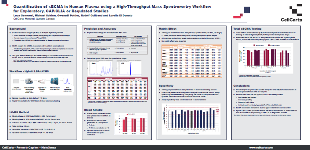
CellCarta’s poster presents a high-throughput hybrid immunoaffinity LC-MRM based method for quantifying soluble BCMA (sBCMA) in human plasma.

The study demonstrates that sBCMA, a marker for multiple myeloma, can be precisely measured using this method, which overcomes interferences from ligands and therapeutic antibodies. The assay shows high sensitivity, a broad linear range, and robustness against interferences, making it suitable for clinical studies and monitoring treatment response in multiple myeloma patients

Quantification of sBCMA in Human Plasma using a High-ThroughputHybrid IP-MRM Based Mass Spectrometry Workflow, As presented at ASMS 2022
sBCMA Plasma Quantification via Hybrid Mass Spectrometry

July 27, 2021

CellCarta’s poster presents a high-throughput hybrid immunoaffinity LC-MRM based method for quantifying soluble BCMA (sBCMA) in human plasma.

The study demonstrates that sBCMA, a marker for multiple myeloma, can be precisely measured using this method, which overcomes interferences from ligands and therapeutic antibodies. The assay shows high sensitivity, a broad linear range, and robustness against interferences, making it suitable for clinical studies and monitoring treatment response in multiple myeloma patients

Quantification of sBCMA in Human Plasma using a High-ThroughputHybrid IP-MRM Based Mass Spectrometry Workflow, As presented at ASMS 2022

Wide Array of Services in Multiple Myeloma

June 21, 2021

DOWNLOAD OUR INFOGRAPHIC

Multiple myeloma infographic

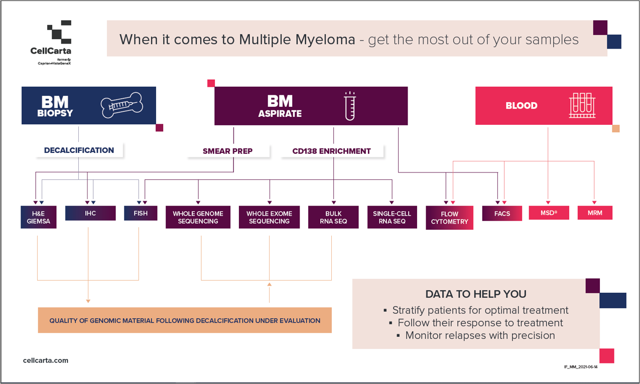
Wide Array of Services in Multiple Myeloma

June 21, 2021

DOWNLOAD OUR INFOGRAPHIC

Multiple myeloma infographic

Effective Biomarker Strategies for Emerging Multiple Myeloma Therapies

June 9, 2021

Section image

Targeting BMCA in multiple myeloma (MM) has leapt into clinical practice, most recently with the approvals of a new antibody drug conjugate (BLENREP, GSK, 2020) and a chimeric antigen receptor T-cell therapy (CAR T, ABECMA, BMS, 2021). While BCMA has shown great promise, several new drugs directed at other targets for treatment of MM have recently been approved or are in development, greatly expanding treatment options in relapsed or refractory settings.

While there is an increasing number of clinical trials investigating drugs targeting BCMA (from 5 in 2015 to >30 in 2019 according to clinicaltrials.gov), other targets have been pursued, including: CD38, HDAC, and SLAMF7.

For each of these targets, multiple therapeutic modalities across small molecules, monoclonal antibodies (mAb), antibody drug conjugates (ADCs), bi-specific antibodies and more recently CD3-directed bi-specifics T-cell engagers (BiTEs) and CAR-T therapies are being investigated. However, each target and drug combination represent a unique translational biomarker challenge with different combinations of mechanisms of action (MOA) and therapeutic pathways. This complexity is something we are keenly focused on at CellCarta where we bring a comprehensive technology approach to addressing biomarker strategies and deliver biomarker data to support clinical programs.

Section image

Biomarkers to stratify MM patients with greater precision

Patient stratification in clinical trials depends on prognostic factors such as disease burden and staging, tumor biology, but also on the expression of the target proteins of the investigational treatments. To enable stratification with greater precision, CellCarta offers multiple assays investigating key biomarkers in CD138 enriched bone marrow cells.

Using bone marrow biopsies, IHC can be performed to further profile the tumor as well as the tumor micro-environment (TME) using key markers such as CD38, CD138, BCMA.

Monitoring immune responses in MM patients using key biomarkers

Flow cytometry allows the direct measurement of cells with abnormal phenotypes associated with disease progression. At CellCarta we deploy off-the-shelf and custom multi-color flow panels to phenotypically characterize a variety of immune subsets acting as key biomarkers across all phases of drug development.

A specific plasma cell panel, including markers such as CD138, CD38, BCMA and PD-L1, was developed to monitor the presence of abnormal plasma cells in both blood and bone marrow aspirate clinical samples. BD TrucountTM tubes can provide both frequencies and absolute counts readouts. Using this panel, important prognostic factors relating to treatments such as BCMA and PD-L1 can be investigated on subsets of interest. For individuals undergoing anti-CD38 therapy, the use of a polyclonal anti-CD38 antibody allows us to detect the receptor. The tumor microenvironment (TME) can also be investigated using flow cytometry and customized panels.

Tracking the course of MM with soluble biomarkers

Soluble blood-based biomarkers have been used to diagnose and monitor the course of MM since the inclusion of serum free light chain (FLC) concentrations in the 2014 IMWG criteria. While FLC assays are a routine part of the MM clinical biomarker analysis, along with the haemato-pathological and flow cytometry workflows, the emergence of BCMA targeted therapies has led to the addition of soluble BCMA concentrations as a critical biomarker. At CellCarta we have developed an off-the-shelf hybrid IP-LC-MRM based assay for the quantification of soluble BCMA in plasma for both diagnostic and prognostic utility. The assay is notably robust to interference from endogenous ligands (APRIL and BAFF), and therapeutic antibodies.

These biomarker strategies are complemented with broader immune monitoring capabilities essential in investigating a patient’s response to treatments. These include cytokine measurement by Meso Scale Discovery (CLIA-validated proinflammatory panel) and broad immune monitoring by flow cytometry. CellCarta’s deep expertise in immunology helps us address all the needs when it comes to MM clinical research.


About the author:

author photo

Nick Dupuis is a Scientific Business Director at CellCarta, specialized in proteomics and biophysical measurement. He has held various roles in industry including as a scientist in LDT/IVD product development and supporting applications and business development for emerging analytical platforms.

author photo

Damien Montamat-Sicotte is a Scientific Business Director at CellCarta, specializing in the flow Cytometry platform. With a PhD in immunology and post-doctoral expertise from various institutions, Damien has profuse experience in managing the processing and analysis of clinical samples by flow cytometry in an immune monitoring context.

Effective Biomarker Strategies for Emerging Multiple Myeloma Therapies

June 9, 2021

Section image

Targeting BMCA in multiple myeloma (MM) has leapt into clinical practice, most recently with the approvals of a new antibody drug conjugate (BLENREP, GSK, 2020) and a chimeric antigen receptor T-cell therapy (CAR T, ABECMA, BMS, 2021). While BCMA has shown great promise, several new drugs directed at other targets for treatment of MM have recently been approved or are in development, greatly expanding treatment options in relapsed or refractory settings.

While there is an increasing number of clinical trials investigating drugs targeting BCMA (from 5 in 2015 to >30 in 2019 according to clinicaltrials.gov), other targets have been pursued, including: CD38, HDAC, and SLAMF7.

For each of these targets, multiple therapeutic modalities across small molecules, monoclonal antibodies (mAb), antibody drug conjugates (ADCs), bi-specific antibodies and more recently CD3-directed bi-specifics T-cell engagers (BiTEs) and CAR-T therapies are being investigated. However, each target and drug combination represent a unique translational biomarker challenge with different combinations of mechanisms of action (MOA) and therapeutic pathways. This complexity is something we are keenly focused on at CellCarta where we bring a comprehensive technology approach to addressing biomarker strategies and deliver biomarker data to support clinical programs.

Section image

Biomarkers to stratify MM patients with greater precision

Patient stratification in clinical trials depends on prognostic factors such as disease burden and staging, tumor biology, but also on the expression of the target proteins of the investigational treatments. To enable stratification with greater precision, CellCarta offers multiple assays investigating key biomarkers in CD138 enriched bone marrow cells.

Using bone marrow biopsies, IHC can be performed to further profile the tumor as well as the tumor micro-environment (TME) using key markers such as CD38, CD138, BCMA.

Monitoring immune responses in MM patients using key biomarkers

Flow cytometry allows the direct measurement of cells with abnormal phenotypes associated with disease progression. At CellCarta we deploy off-the-shelf and custom multi-color flow panels to phenotypically characterize a variety of immune subsets acting as key biomarkers across all phases of drug development.

A specific plasma cell panel, including markers such as CD138, CD38, BCMA and PD-L1, was developed to monitor the presence of abnormal plasma cells in both blood and bone marrow aspirate clinical samples. BD TrucountTM tubes can provide both frequencies and absolute counts readouts. Using this panel, important prognostic factors relating to treatments such as BCMA and PD-L1 can be investigated on subsets of interest. For individuals undergoing anti-CD38 therapy, the use of a polyclonal anti-CD38 antibody allows us to detect the receptor. The tumor microenvironment (TME) can also be investigated using flow cytometry and customized panels.

Tracking the course of MM with soluble biomarkers

Soluble blood-based biomarkers have been used to diagnose and monitor the course of MM since the inclusion of serum free light chain (FLC) concentrations in the 2014 IMWG criteria. While FLC assays are a routine part of the MM clinical biomarker analysis, along with the haemato-pathological and flow cytometry workflows, the emergence of BCMA targeted therapies has led to the addition of soluble BCMA concentrations as a critical biomarker. At CellCarta we have developed an off-the-shelf hybrid IP-LC-MRM based assay for the quantification of soluble BCMA in plasma for both diagnostic and prognostic utility. The assay is notably robust to interference from endogenous ligands (APRIL and BAFF), and therapeutic antibodies.

These biomarker strategies are complemented with broader immune monitoring capabilities essential in investigating a patient’s response to treatments. These include cytokine measurement by Meso Scale Discovery (CLIA-validated proinflammatory panel) and broad immune monitoring by flow cytometry. CellCarta’s deep expertise in immunology helps us address all the needs when it comes to MM clinical research.


About the author:

author photo

Nick Dupuis is a Scientific Business Director at CellCarta, specialized in proteomics and biophysical measurement. He has held various roles in industry including as a scientist in LDT/IVD product development and supporting applications and business development for emerging analytical platforms.

author photo

Damien Montamat-Sicotte is a Scientific Business Director at CellCarta, specializing in the flow Cytometry platform. With a PhD in immunology and post-doctoral expertise from various institutions, Damien has profuse experience in managing the processing and analysis of clinical samples by flow cytometry in an immune monitoring context.

Validated Flow Cytometry for Monitoring Multiple Myeloma

November 9, 2020

CellCarta’s poster presents a validated flow cytometry panel for the clinical monitoring of multiple myeloma (MM) patients. The MM Counting Panel detects and phenotypes malignant and non-malignant plasma cells, T cells, B cells, monocytes, and NK cells using fluorochrome-conjugated antibodies.

Key Features of the MM Counting Panel:

  • Detection and Enumeration: Accurately enumerates immune populations in bone marrow aspirates (BMA) and peripheral blood (PB).
  • Phenotyping: Identifies specific MM markers, including BCMA, using a surface marker profile (CD19-CD56+BCMA+).
  • Intra- and Inter-assay Precision: Demonstrated high precision with PB samples from both healthy and MM donors, ensuring reliability of results.
  • Sample Stability: Maintained stability and met acceptance criteria for key markers across different temperature conditions and time points.
  • Clinical Validation: Validated with clinical samples from the NCT03761108 trial, showing different BCMA expression patterns among subjects.
  • sBCMA measurement by mass spectrometry can be used as a complementary method for biomarker monitoring.

Assay Development and Characterization:

  • Sample Collection and Preparation: PB and BMA samples were obtained from healthy and MM donors, processed in Cyto-Chex® blood collection tubes, and analyzed using BD TruCount™ tubes.
  • Gating Hierarchy: Established for the identification of malignant plasma cells, with a specific focus on CD45+CD19-CD56+BCMA+ populations.

Conclusion: The MM Counting Panel, validated for clinical use, allows for robust and reliable monitoring of multiple myeloma patients. This comprehensive approach combines flow cytometry and mass spectrometry to enhance the detection of key biomarkers, supporting the development of novel therapies and optimizing treatment outcomes.

Multiple Myeloma Flow Cytometry Panel Validated for Clinical Monitoring of Patients

As presented in SITC 2020

Capion_Poster_SITC_BCMAflow

Validated Flow Cytometry for Monitoring Multiple Myeloma

November 9, 2020

CellCarta’s poster presents a validated flow cytometry panel for the clinical monitoring of multiple myeloma (MM) patients. The MM Counting Panel detects and phenotypes malignant and non-malignant plasma cells, T cells, B cells, monocytes, and NK cells using fluorochrome-conjugated antibodies.

Key Features of the MM Counting Panel:

  • Detection and Enumeration: Accurately enumerates immune populations in bone marrow aspirates (BMA) and peripheral blood (PB).
  • Phenotyping: Identifies specific MM markers, including BCMA, using a surface marker profile (CD19-CD56+BCMA+).
  • Intra- and Inter-assay Precision: Demonstrated high precision with PB samples from both healthy and MM donors, ensuring reliability of results.
  • Sample Stability: Maintained stability and met acceptance criteria for key markers across different temperature conditions and time points.
  • Clinical Validation: Validated with clinical samples from the NCT03761108 trial, showing different BCMA expression patterns among subjects.
  • sBCMA measurement by mass spectrometry can be used as a complementary method for biomarker monitoring.

Assay Development and Characterization:

  • Sample Collection and Preparation: PB and BMA samples were obtained from healthy and MM donors, processed in Cyto-Chex® blood collection tubes, and analyzed using BD TruCount™ tubes.
  • Gating Hierarchy: Established for the identification of malignant plasma cells, with a specific focus on CD45+CD19-CD56+BCMA+ populations.

Conclusion: The MM Counting Panel, validated for clinical use, allows for robust and reliable monitoring of multiple myeloma patients. This comprehensive approach combines flow cytometry and mass spectrometry to enhance the detection of key biomarkers, supporting the development of novel therapies and optimizing treatment outcomes.

Multiple Myeloma Flow Cytometry Panel Validated for Clinical Monitoring of Patients

As presented in SITC 2020

Capion_Poster_SITC_BCMAflow

Antigen Presentation: A Critical Step in Immune Therapy Dev.

June 10, 2020

In this webinar you will learn:

  • Successful antigen presentation is required to initiate an immune response
  • The most physiologically relevant way to measure the full breadth of antigen presentation is by direct observation of presented peptides using mass spectrometry
  • The quality and robustness of the mass spectrometry data generation is therefore a critical element

What is antigen presentation?

Antigen presentation is essential for development of a robust immune response.

Binding of an MHC-peptide complex by a T cell receptor is the initial and critical step for starting a new adaptive immune response or for regulating an ongoing one. Identification of effectively presented antigens is therefore a defining element for any successful immune therapy.

Antigen presentation can occur through MHC class I, class II, or non-classical MHC proteins. Each protein complex has characteristic tissue expression, type of antigen peptide presented, and role in the immune response.

Most lymphocytes including all of those with antigen presenting functions can express both MHC I and MHC II, and involvement of both MHC I- and MHC II-restricted responses is considered essential for a full immune response.

Non-classical MHC such as HLA-E and HLA-G are characterized by either a relatively restricted tissue distribution or antigen presentation repertoire and have specific roles to play in the establishment of robust immunity.

Direct observation of presented peptides by mass spectrometry has become required for novel epitope discovery. This approach is the most physiologically relevant way to detect the modified and unmodified peptides presented by any type of MHC.

Additionally, mass spectrometry can be used to quantify the peptide presentation to ensure that attractive targets are sufficiently expressed to trigger an immune reaction. The limiting factor has now become the quality of the mass spectrometry data generated.

This presentation will show examples of direct identification of novel presented peptides as well as survey the requirements for the industrialization of antigen presentation mass spectrometry to generate robust, reliable and quantitative measurements.

With an understanding that antigen presentation is required to initiate an immune response, we will discuss how to use this information in therapeutics development. Designing a good immune therapy requires that the antigen is specific to the targeted pathology, as well as immunogenic.

Those factors are especially relevant for modern immune therapies that seek to engage the host response as part of the complete therapy. Antigen presentation by mass spectrometry should therefore be embedded in a broader, integrated, characterization of host immunity.

Specific characterizations include evaluation of wider host antigen presentation to assess target specificity, monitoring the status of the host microenvironment, as well as host immune exhaustion before and after antigen dosing to better understand the capacity to respond to the therapy.

This presentation will depict case studies addressing each of these components of successful immune therapy candidates that start with successful antigen presentation.

Antigen presentation is the initial critical element, but for successful immune therapy development, antigen presentation should be embedded in an integrated assessment of host immunity

Speaker: Eustache Paramithiotis PhD, Vice-President, Research & Development, Caprion Biosciences Inc.

Antigen Presentation: A Critical Step in Immune Therapy Dev.

June 10, 2020

In this webinar you will learn:

  • Successful antigen presentation is required to initiate an immune response
  • The most physiologically relevant way to measure the full breadth of antigen presentation is by direct observation of presented peptides using mass spectrometry
  • The quality and robustness of the mass spectrometry data generation is therefore a critical element

What is antigen presentation?

Antigen presentation is essential for development of a robust immune response.

Binding of an MHC-peptide complex by a T cell receptor is the initial and critical step for starting a new adaptive immune response or for regulating an ongoing one. Identification of effectively presented antigens is therefore a defining element for any successful immune therapy.

Antigen presentation can occur through MHC class I, class II, or non-classical MHC proteins. Each protein complex has characteristic tissue expression, type of antigen peptide presented, and role in the immune response.

Most lymphocytes including all of those with antigen presenting functions can express both MHC I and MHC II, and involvement of both MHC I- and MHC II-restricted responses is considered essential for a full immune response.

Non-classical MHC such as HLA-E and HLA-G are characterized by either a relatively restricted tissue distribution or antigen presentation repertoire and have specific roles to play in the establishment of robust immunity.

Direct observation of presented peptides by mass spectrometry has become required for novel epitope discovery. This approach is the most physiologically relevant way to detect the modified and unmodified peptides presented by any type of MHC.

Additionally, mass spectrometry can be used to quantify the peptide presentation to ensure that attractive targets are sufficiently expressed to trigger an immune reaction. The limiting factor has now become the quality of the mass spectrometry data generated.

This presentation will show examples of direct identification of novel presented peptides as well as survey the requirements for the industrialization of antigen presentation mass spectrometry to generate robust, reliable and quantitative measurements.

With an understanding that antigen presentation is required to initiate an immune response, we will discuss how to use this information in therapeutics development. Designing a good immune therapy requires that the antigen is specific to the targeted pathology, as well as immunogenic.

Those factors are especially relevant for modern immune therapies that seek to engage the host response as part of the complete therapy. Antigen presentation by mass spectrometry should therefore be embedded in a broader, integrated, characterization of host immunity.

Specific characterizations include evaluation of wider host antigen presentation to assess target specificity, monitoring the status of the host microenvironment, as well as host immune exhaustion before and after antigen dosing to better understand the capacity to respond to the therapy.

This presentation will depict case studies addressing each of these components of successful immune therapy candidates that start with successful antigen presentation.

Antigen presentation is the initial critical element, but for successful immune therapy development, antigen presentation should be embedded in an integrated assessment of host immunity

Speaker: Eustache Paramithiotis PhD, Vice-President, Research & Development, Caprion Biosciences Inc.