High-quality immunofluorescence (IF) and brightfield immunohistochemistry (IHC) services supported by our pathology team and imaging scientists.
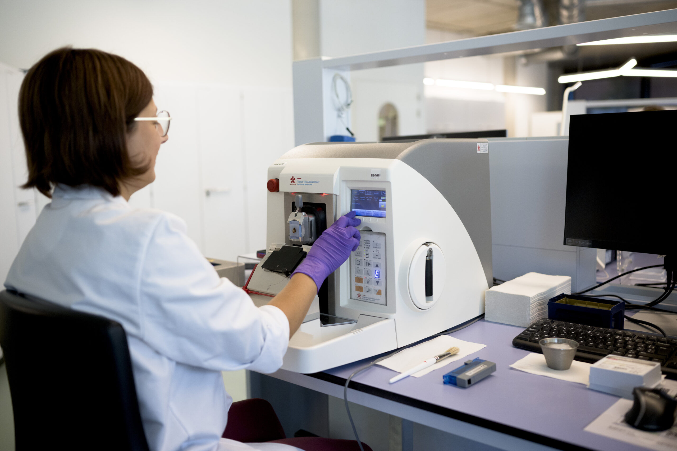
At CellCarta, we leverage our expertise and experience in IHC to obtain deep insight into the abundance, distribution, and localization of your program’s target antigens in tissues and cells.
We offer customized assay development services tailored to fit your biomarker strategy, study phase, and assay use case. Singleplex, multiplex, and hyperplex assays can be developed in combination with analysis methods to obtain relevant and accurate data when clinical samples are limited or scarce.
Depending on your study’s needs, we offer a comprehensive range of validated off-the-shelf (OTS) assays for any species of interest.
With CAP/CLIA accreditations and ISO-certified workflows, our teams always adhere to the highest standard of quality during development.
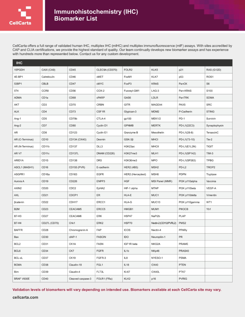
View our in-depth IHC biomarker list here. Our team has experience with hundreds more targets than what’s listed; contact us to discuss your requirements further.
End-to-end IHC development services are available, starting from the construction of prototype assays through to CDx level validation. Our team’s strategic selection of primary antibodies and control tissues, advanced staining and scanning instruments, and standardized protocols ensures optimal panel development.
Each program is unique, so we ensure that our assay development approach is flexible and tailored to your study’s requirements.
Commitment to quality and consistency is central to CellCarta’s global assay development strategy. Global immunohistochemistry services are delivered within a single quality framework and guided by standardized SOPs.
CellCarta assay development scientists and pathologists work with clients and vendors to ensure accurate, robust and reproducible assay construction and validation according to your program’s requirements.
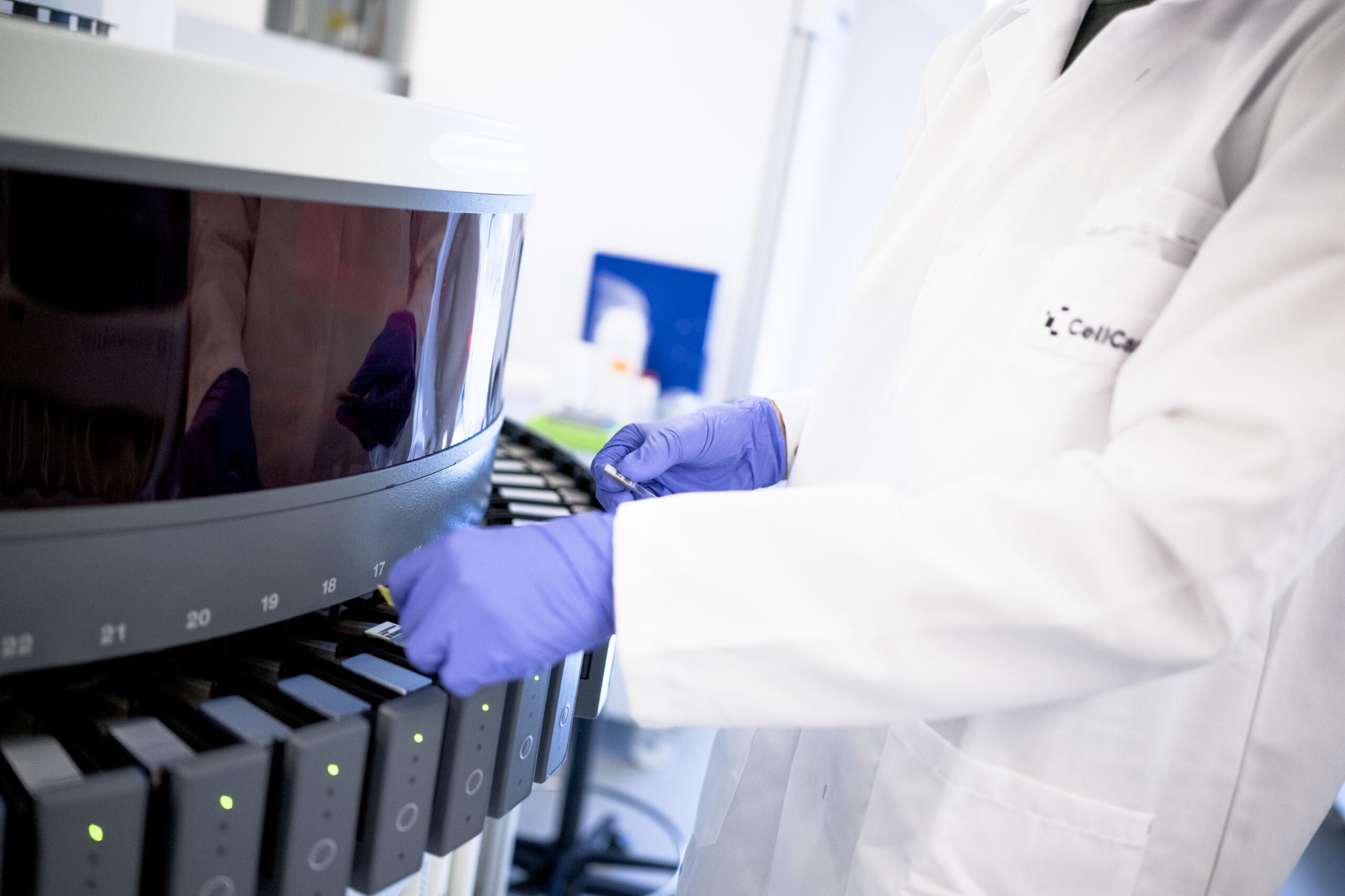
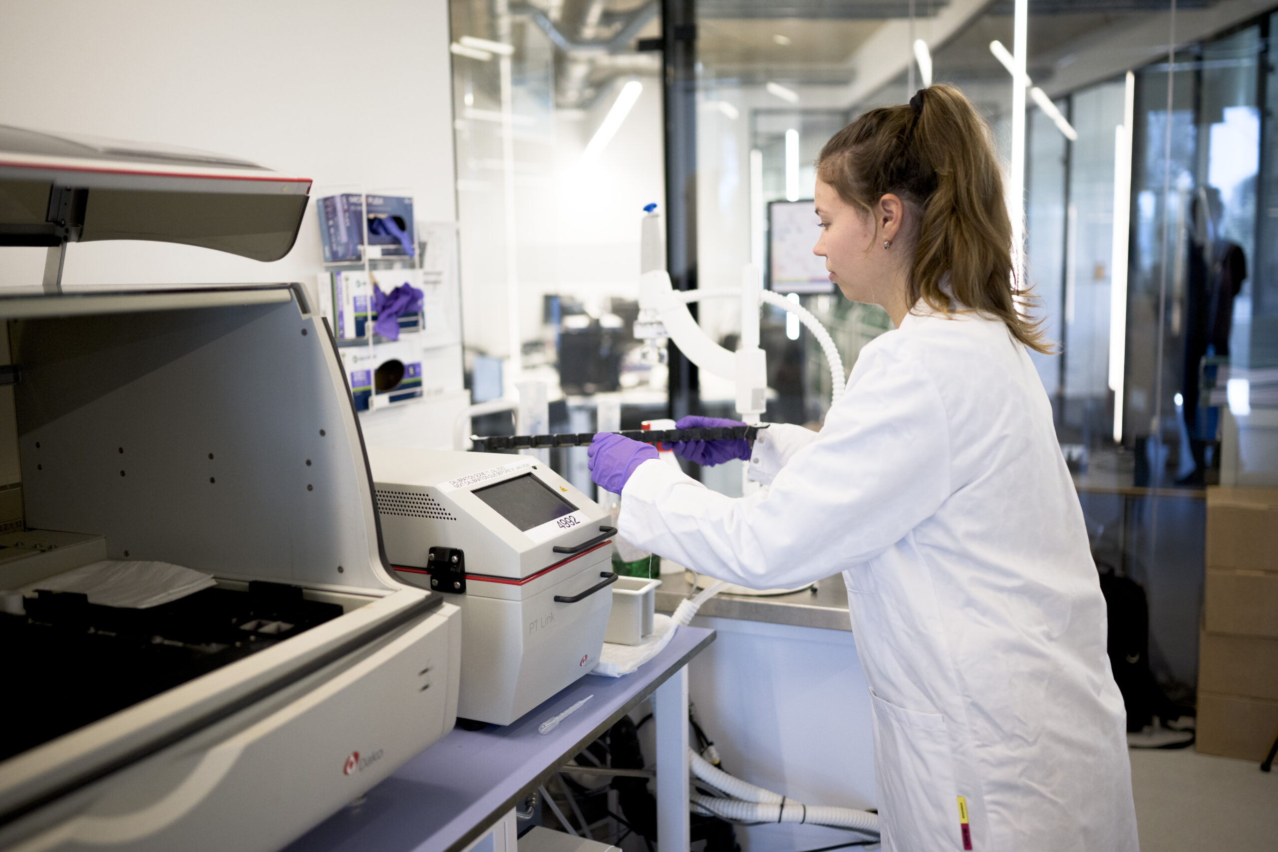
Assays are developed on the same automated platforms used for clinical testing, which eliminates the need for cross-validation, minimizes variability, and accelerates trial timelines. We are platform agnostic and maintain a global suite of automated stainers, including:
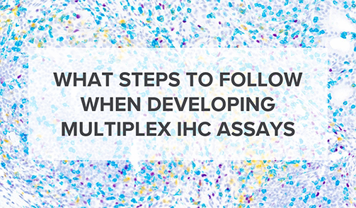
We can develop multiplex (up to 6 targets) and hyperplex (up to 40 targets) assays using standardized workflows with advanced fluorescent and brightfield reagents. Key advantages include:
Depending on your study’s needs, we offer a comprehensive range of validated off-the-shelf (OTS) assays for any species of interest.
With CAP/CLIA accreditations and ISO-certified workflows, our teams always adhere to the highest standard of quality during development.
View our in-depth IHC biomarker list here. Our team has experience with hundreds more targets than what’s listed; contact us to discuss your requirements further.
End-to-end IHC development services are available, starting from the construction of prototype assays through to CDx level validation. Our team’s strategic selection of primary antibodies and control tissues, advanced staining and scanning instruments, and standardized protocols ensures optimal panel development.
Each program is unique, so we ensure that our assay development approach is flexible and tailored to your study’s requirements.
Commitment to quality and consistency is central to CellCarta’s global assay development strategy. Global immunohistochemistry services are delivered within a single quality framework and guided by standardized SOPs.
CellCarta assay development scientists and pathologists work with clients and vendors to ensure accurate, robust and reproducible assay construction and validation according to your program’s requirements.
Assays are developed on the same automated platforms used for clinical testing, which eliminates the need for cross-validation, minimizes variability, and accelerates trial timelines. We are platform agnostic and maintain a global suite of automated stainers, including:
We can develop multiplex (up to 6 targets) and hyperplex (up to 40 targets) assays using standardized workflows with advanced fluorescent and brightfield reagents. Key advantages include:
Every IHC assay developed at CellCarta is paired with one or more analysis methods, including pathologist scoring, CellCarta-developed image analysis, or third-party algorithms. Once assay analytes are defined, all data capture, storage, and reporting are fully mapped, validated, and quality checked.
A biotech team needed two custom 6-plex mIF assays developed and 87 clinical FFPE samples tested quickly before a conference submission deadline. CellCarta's Lake Forest laboratory delivered over 9,000 data points ahead of schedule, with results presented in ready-to-use formats for easy presentation.
Singleplex to hyperplex assays backed by our pathologists and rigorous quality workflows.
Platform Agnostic
Validated Assay Offerings
Pathologist Oversight
Complex Biomarker Expertise
Designed for Clinical Use
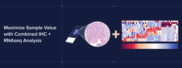
Achieve a fuller understanding of your target cells and biomarkers by combining our IHC services with our other offerings.
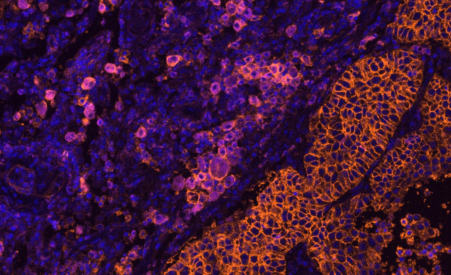
Immunohistochemistry uses enzyme-based chromogenic detection to visualize protein targets in tissue sections, producing brightfield or immunofluorescent images that can be evaluated by pathologists using standard light microscopy. Immunofluorescence uses fluorescent-labeled antibodies to detect one or more targets simultaneously on a single slide, enabling multiplexed protein co-localization and spatial analysis. Both approaches are available at CellCarta, spanning singleplex IHC to hyperplex IF assays covering up to 40 targets. The choice of platform depends on the number of targets, tissue availability, and the intended analytical output.
CellCarta provides end-to-end immunohistochemistry services, from early assay development through to clinical trial testing and companion diagnostic support. Services span singleplex IHC, multiplex IHC, multiplex immunofluorescence, and hyperplex IF. Assay development covers antibody selection, platform selection, control tissue selection, and experimental condition optimization.
CellCarta is platform agnostic and operates a global suite of automated stainers across multiple manufacturers. Assays are developed on the same automated staining platforms used for clinical sample testing, which eliminates the need for cross-validation, minimizes variability, and reduces the time between assay validation and clinical trial testing. Available platforms include Dako Autostainer Link48 (with PT-modules), Dako Omnis, Ventana Benchmark Ultra, Ventana Benchmark Ultra Plus, Ventana Discovery Ultra, Leica Bond Rx, Leica Bond III, and Lunaphore COMET.
Multiplex IHC assays allow multiple protein targets to be detected on a single tissue slide, generating in-depth biomarker data even when sample volume is limited or scarce. Key advantages include accurate tissue and cellular segmentation for image analysis, single-view multianalyte data integration for mechanistic pathway insights, and elucidation of the mechanism of action of candidate therapeutics. AI-supported pathologist scoring is available for all multiplex and hyperplex outputs. Every result is reviewed by a board-certified pathologist before delivery, ensuring scientific rigor and data quality throughout the workflow.
CellCarta's digital pathology infrastructure supports whole slide imaging and AI-assisted image analysis for both brightfield and fluorescent slides. Available scanners include the Roche Ventana DP600, Leica Aperio AT Turbo, and Akoya PhenoImager HT, which supports multispectral fluorescence imaging for up to nine color channels. Image analysis workflows are all 21 CFR Part 11 compliant, supporting data integrity requirements for regulated clinical studies and IND-enabling programs.