Longitudinal tracking of cell proliferation dynamics and cell cycling using flow cytometry.
CellCarta’s flow cytometry expertise enables seamless understanding of your cells and their dynamic behavior. We tailor our workflow to your study’s specifications, with multiple cell proliferation assays available for use in both tissue and liquid biopsy samples.
Our analysis doesn’t stop there. Our workflow can incorporate additional surface and intracellular markers into our proliferation workflow for in-depth insight into your cell subsets.
We use flow cytometry to phenotypically and functionally profile distinct immune cell subsets from whole blood, peripheral blood mononuclear cells (PBMCs), bone marrow aspirates (BMA), cerebrospinal fluid (CSF), and dissociated tissue samples, measuring both proliferative activity and cell surface identity in a single assay.
Our qualified CFSE (carboxyfluorescein succinimidyl ester) assay quantifies proliferation by tracking dye dilution across successive cell divisions. Suited for measuring cellular response to clinical treatment and in vitro stimulations, it delivers a reliable proliferation index for each cell subset analyzed.
Ki-67 is used as an early activation marker to identify cells that enter a new cell cycle. Expressed across all active cell phases (G1, S, G2, and M) yet absent from resting cells (G0), Ki-67 provides a direct window into cycling activity across your cellular subsets of interest.
Bromodeoxyuridine (BrdU)-based DNA synthesis measurement captures newly proliferating cells through their incorporation into freshly synthesized DNA. This technique complements dye dilution and protein-based approaches when direct DNA synthesis quantification is required.
We use flow cytometry to phenotypically and functionally profile distinct immune cell subsets from whole blood, peripheral blood mononuclear cells (PBMCs), bone marrow aspirates (BMA), cerebrospinal fluid (CSF), and dissociated tissue samples, measuring both proliferative activity and cell surface identity in a single assay.
Our qualified CFSE (carboxyfluorescein succinimidyl ester) assay quantifies proliferation by tracking dye dilution across successive cell divisions. Suited for measuring cellular response to clinical treatment and in vitro stimulations, it delivers a reliable proliferation index for each cell subset analyzed.
Ki-67 is used as an early activation marker to identify cells that enter a new cell cycle. Expressed across all active cell phases (G1, S, G2, and M) yet absent from resting cells (G0), Ki-67 provides a direct window into cycling activity across your cellular subsets of interest.
Bromodeoxyuridine (BrdU)-based DNA synthesis measurement captures newly proliferating cells through their incorporation into freshly synthesized DNA. This technique complements dye dilution and protein-based approaches when direct DNA synthesis quantification is required.
Our cell proliferation assays are designed for the clinical space. Their uses include assessing immune cell functionality and responsiveness, determining the proliferation index of cell subsets, identifying actively proliferating cells, and tracking treatment response and disease course over time.
Expert implementation of cell proliferation assays for rich cellular insights.
Validated for Clinical Use
Broad Profiling Options
Global Execution
Multi-Technique Expertise
Cross-Validated Protocols
Explore the avenues through which flow cytometry platforms and analysis can benefit your study.
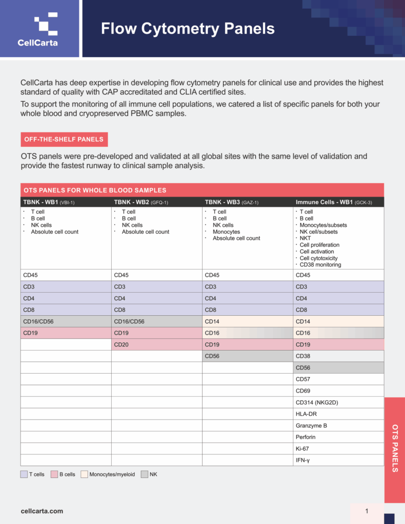
Characterize complex immune cell subsets using off-the-shelf or fully customized panels.
Detect cytokine production at the single-cell level to profile immune function.
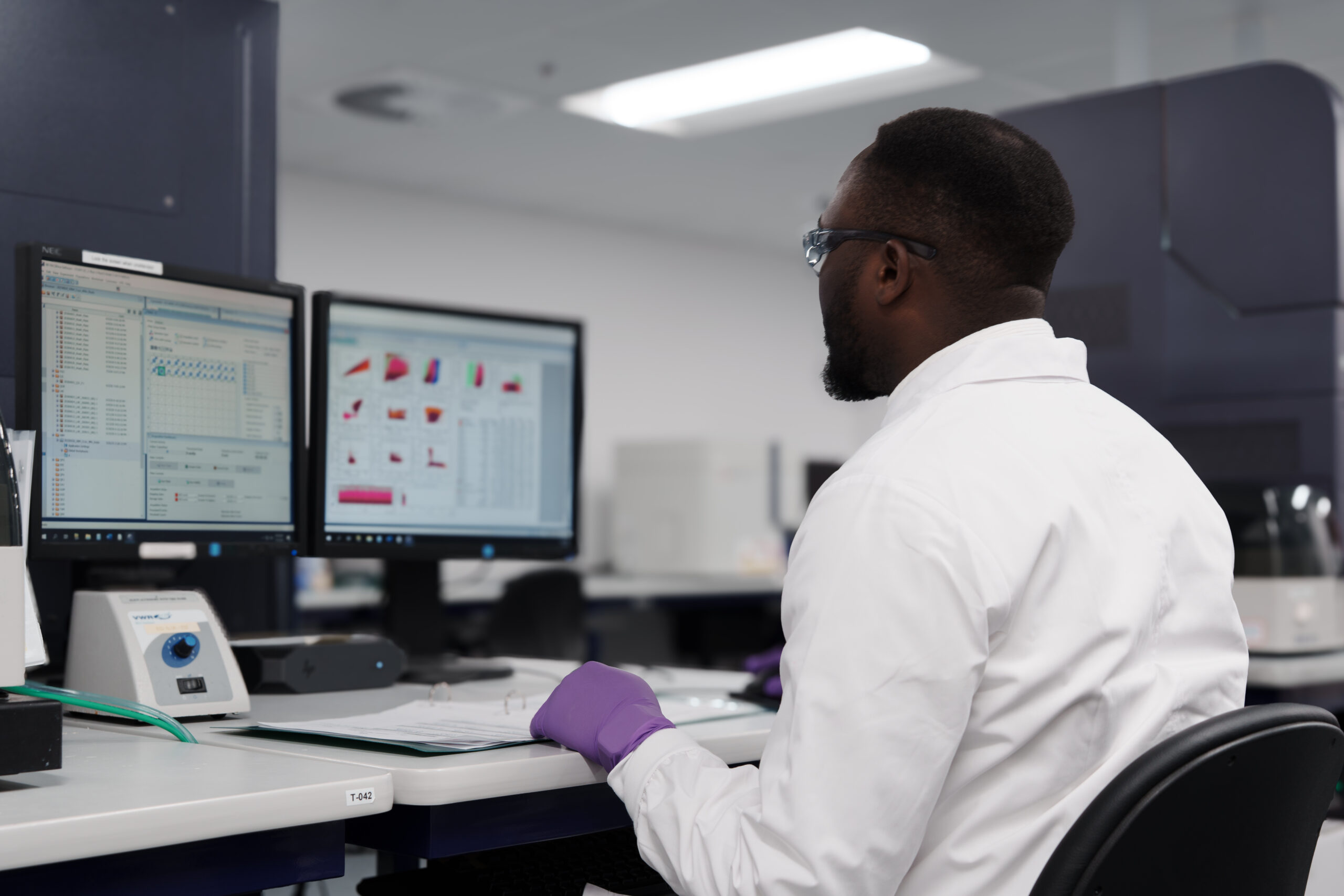
Advanced flow cytometry analysis and data visualization with incredible speed.

Cell proliferation assays provide direct, quantitative insight into how immune cells respond to treatment. By measuring cell proliferation through a flow cytometry-based workflow, your program can determine the proliferation index of a specific cell subset, identify which populations are actively dividing, and track changes in cell cycling behavior across the course of treatment or disease.
A key benefit is the ability to compare immune cell responsiveness across diseased and healthy individuals within the same study. When combined with additional cell surface and intracellular markers, cell proliferation assays deliver complete, broad insights about specific cell subsets, going beyond a simple cell count to reveal phenotypic and functional insights.
We use multiple techniques to track cells during the proliferation process: BrdU, which measures DNA synthesis by incorporating into newly synthesized DNA; CFSE, which quantifies proliferation by tracking dye dilution across generations; and labelled cell cycle-associated proteins (e.g., Ki-67 and PCNA - proliferating cell nuclear antigen) to monitor cellular dynamics. Each technique can be selected based on the specific requirements of your program.
Our cell proliferation assays can accommodate a wide range of clinical sample types, including whole blood, PBMCs (fresh and cryopreserved), tissue and tumor samples, BMA, fine-needle aspiration biopsies, CSF, and cell lines. Sample type suitability can be confirmed during initial consultations with our scientific experts.
CFSE (carboxyfluorescein succinimidyl ester) tracks dye dilution overtime to quantify cell proliferation. As a labeled cell divides, the fluorescent signal is distributed equally between daughter cells, allowing each successive generation to be distinguished. CellCarta offers a qualified CFSE assay suitable for measuring cellular proliferation in response to clinical treatment and in vitro stimulations.
Ki-67 is used as an early activation marker to identify cells that have entered a new cell cycle. The nuclear antigen Ki-67 is expressed across all active phases of the cell cycle (G1, S, G2, M) and is absent from resting cells in G0. CellCarta combines Ki-67 with additional cell surface and intracellular markers to provide broad, functional insights across specific cell subsets.
CellCarta has demonstrated inter-site consistency through a multi-site instrument alignment study that standardized more than 10 assays across five global laboratories simultaneously. The study achieved greater than 80% of readouts with a coefficient of variation (CV) below 30% when comparing reference and receiving laboratories. Lyophilized reference beads were shipped to all sites simultaneously and remain stable at room temperature for up to 18 months, reducing logistical overhead. All cell proliferation assays are performed in a GCLP environment across CAP-accredited and CLIA-certified laboratories.