Flow cytometry-based assays for an in-depth understanding of cytokine expression at the single-cell level.

Cytokine expression reveals key details regarding cell responses, differentiation, and signaling events that can help guide therapeutic development.
At CellCarta, we use flow cytometry for intracellular cytokine staining (ICS) assays to quantify cytokine-producing cells, enabling the functional profiling of immune cell subsets.
Our ICS assays have immense clinical utility and can be readily implemented into clinical trials across many fields, including oncology, autoimmunity, and vaccine development.
Our intracellular cytokine staining assays by flow cytometry use fluorescent antibodies to detect intracellular cytokine expression at the single-cell level.
Unlike bulk cytokine measurement techniques, this approach measures the percentage of cytokine-producing cells, assesses phenotypic changes, and identifies the exact cellular subpopulation expressing the cytokines of interest within a single assay run.
CellCarta cytokine assay services are compatible with a vast range of biological matrices. These clinical sample types include fresh whole blood, cryopreserved PBMCs, and tumor-infiltrating lymphocytes (TILs).
Regardless of the stimulation type used —whether proteins, peptides, or antigen-presenting cells — we provide accurate profiling of cellular subsets across all supported sample matrices.
Our flow cytometry team can develop and validate ICS assays that are suited for your study’s specifications. With decades of experience backing us, we carefully optimize your assay’s parameters to guarantee reliable results.
Regardless of your study’s setup, matrices, and endpoint, we can readily implement an assay for tailored insights into cytokine production.
Our ICS assays enable the simultaneous investigation of intracellular and surface markers within individual cells. HLA class I multimers can be incorporated, allowing for an in-depth understanding of the immune landscape.
Our team can leverage our bioinformatics capabilities and CellEngine software, as well as complementary assays such as CFSE and ELISpot, to deliver a complete profile of antigen-specific responses.
CellCarta's ICS assays support high-level multiplexing, with 16-color panels for conventional cytometry and 30+ color panels for spectral flow cytometry or mass cytometry readily available.
We can simultaneously measure multiple cytokines and surface phenotype markers within a single cell, including assessment of polyfunctionality.
Our intracellular cytokine staining assays by flow cytometry use fluorescent antibodies to detect intracellular cytokine expression at the single-cell level.
Unlike bulk cytokine measurement techniques, this approach measures the percentage of cytokine-producing cells, assesses phenotypic changes, and identifies the exact cellular subpopulation expressing the cytokines of interest within a single assay run.
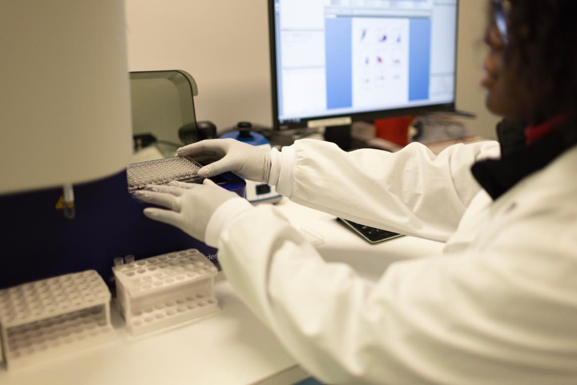
CellCarta cytokine assay services are compatible with a vast range of biological matrices. These clinical sample types include fresh whole blood, cryopreserved PBMCs, and tumor-infiltrating lymphocytes (TILs).
Regardless of the stimulation type used —whether proteins, peptides, or antigen-presenting cells — we provide accurate profiling of cellular subsets across all supported sample matrices.
Our flow cytometry team can develop and validate ICS assays that are suited for your study’s specifications. With decades of experience backing us, we carefully optimize your assay’s parameters to guarantee reliable results.
Regardless of your study’s setup, matrices, and endpoint, we can readily implement an assay for tailored insights into cytokine production.
Our ICS assays enable the simultaneous investigation of intracellular and surface markers within individual cells. HLA class I multimers can be incorporated, allowing for an in-depth understanding of the immune landscape.
Our team can leverage our bioinformatics capabilities and CellEngine software, as well as complementary assays such as CFSE and ELISpot, to deliver a complete profile of antigen-specific responses.
CellCarta's ICS assays support high-level multiplexing, with 16-color panels for conventional cytometry and 30+ color panels for spectral flow cytometry or mass cytometry readily available.
We can simultaneously measure multiple cytokines and surface phenotype markers within a single cell, including assessment of polyfunctionality.

CellCarta’s ICS assays can be used for many applications, such as identifying antigen-specific T cell frequency, determining the phenotype of responding cell populations, analyzing functional subset distribution, and characterizing drug-induced effects on immune functions ex vivo.
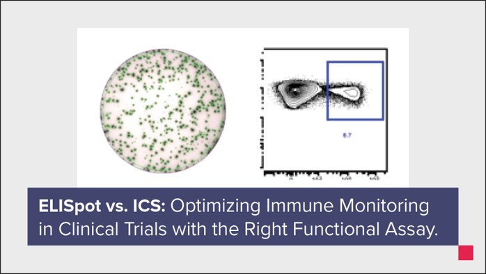
Knowing when to use ELISpot versus ICS to evaluate immune response, particularly for T cells, can be tricky. Our scientists outline key considerations for both assays that serve to optimize your immune monitoring workflow based on your study’s specifications.
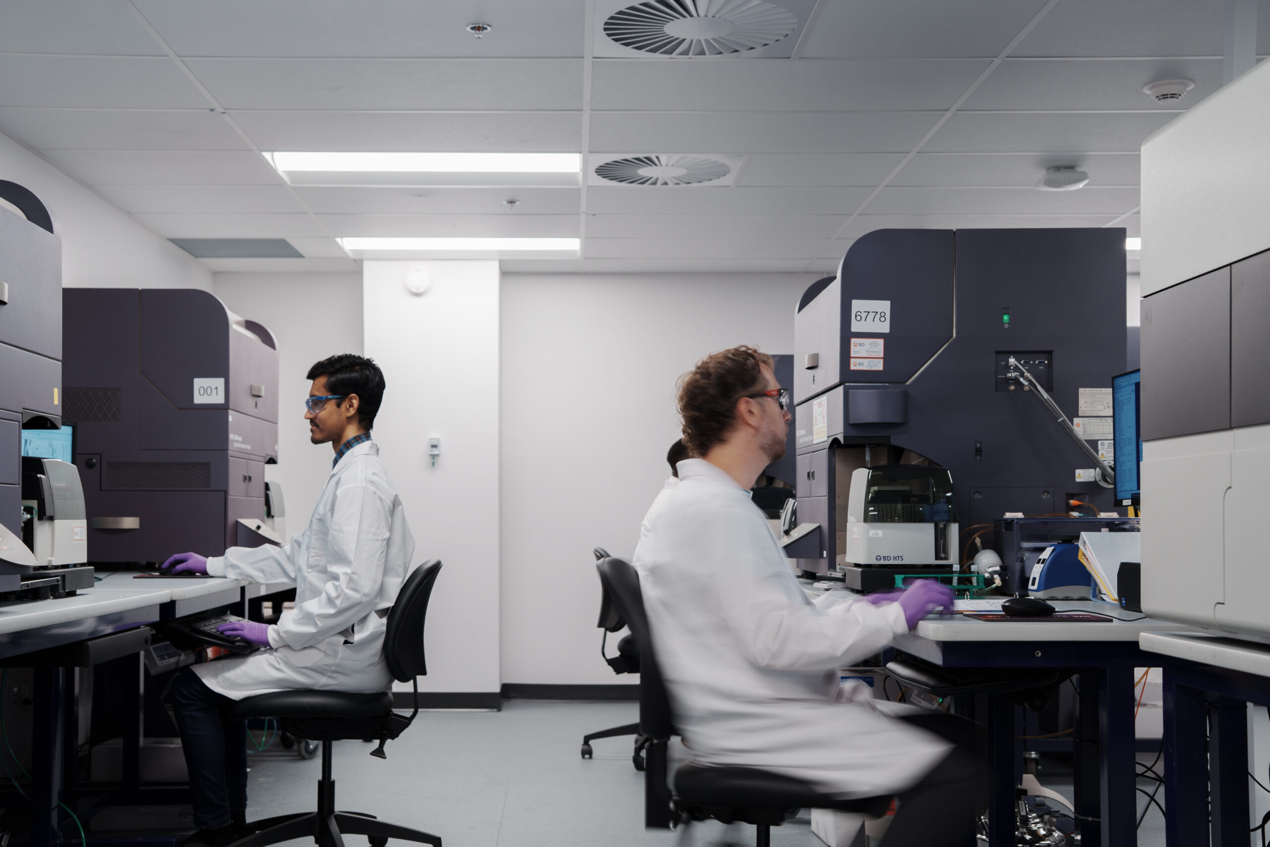
Decades of immune monitoring experience, applied to every ICS program.
Single-Cell Resolution
Multi-Modal Capabilities
Broad Sample Support
High Sensitivity
Custom Assay Design
Enhance the value of your ICS assays by incorporating CellCarta’s related services into your workflow.
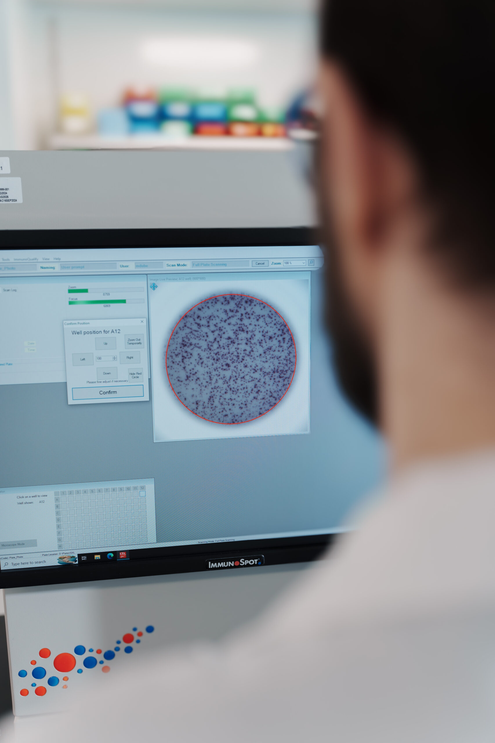
Characterize single-cell cytokine secretion with validated ELISpot and FluoroSpot assays.
Assess immune cell division and cycling to evaluate treatment response and immune activation.
Clinical and bioanalytical expertise to extract biological insights from complex datasets.

ICS uses fluorescent antibodies to detect cytokines trapped inside cells following stimulation. After stimulation with a protein, peptide, or antigen-presenting cell, a secretion inhibitor is added to prevent cytokine release. Cells are then fixed, permeabilized, and stained with fluorescent antibody-conjugated anti-cytokine antibodies and phenotypic markers, allowing simultaneous detection of cytokine production and cell identity by flow cytometry.
CellCarta performs intracellular cytokine staining on fresh whole blood, cryopreserved peripheral blood mononuclear cells (PBMCs), and tumor-infiltrating lymphocytes (TILs). Regardless of the type of stimulation used, the assay delivers accurate profiling of cellular subsets within a sample.
ICS results are expressed as the count or percentage of cytokine-secreting cells within a defined subset. For example, we can measure the percentage of IFN-γ-producing CD8+ T-cells within total CD8+ T-cells. Results can also be expressed as relative cytokine expression levels using median fluorescence intensities (MFI), providing a quantitative measure of how much cytokine each responding cell is producing. CellCarta's bioinformatics team can support downstream analysis of these datasets using CellEngine®.
ICS assay services are best suited to programs requiring detailed phenotypic information on responding cell populations, including distinguishing CD4 from CD8 responses, tracking memory subsets, or assessing polyfunctionality across multiple cytokines simultaneously. ICS is particularly valuable in trials where the immune response may vary based on disease heterogeneity or treatment phase. However, ELISpot remains the preferred choice for focused single-cytokine measurement in well-characterized systems.
Yes. CellCarta supports combined ICS and ELISpot strategies within a single clinical program. ELISpot can be used first as a screening tool to identify which samples warrant deeper investigation. Then a comprehensive intracellular cytokine staining panel is deployed on those selected samples to characterize polyfunctionality and responding cell phenotype in full detail. This two-step approach maximizes scientific return on available sample.