Digital pathology services spanning whole slide scanning, multiplex imaging, and AI-powered image analysis underpinned by nearly two decades of experience.
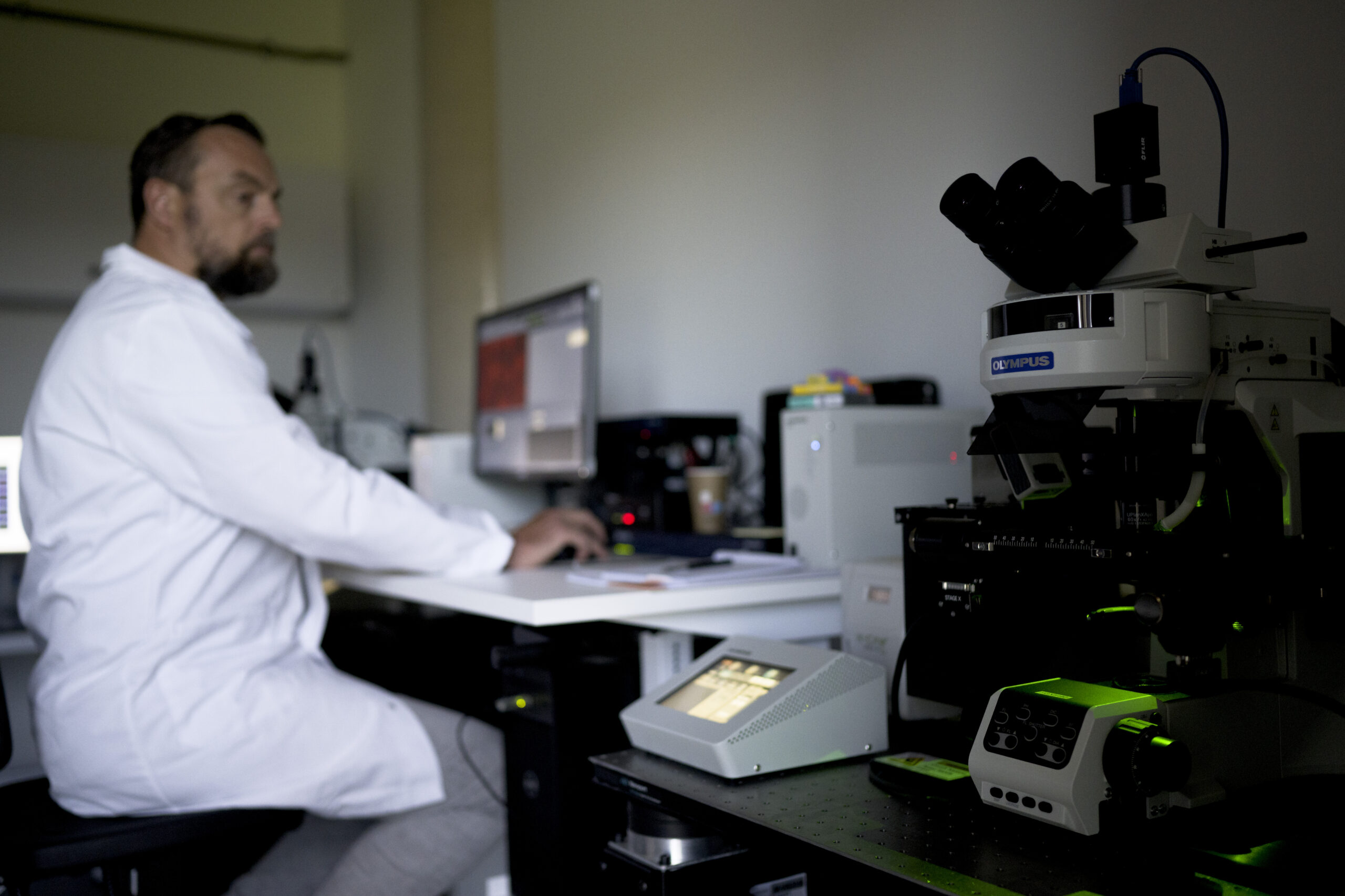
Extract rich, reproducible data from tissue samples with digital pathology solutions built for clinical trial deployment, backed by our team of dedicated in-house pathologists and data scientists.
CellCarta has the foundational structure and expertise needed to adapt to the digital requirements of your program. With decades of experience, we have designed, validated and optimized a robust, centralized cloud-based infrastructure for global deployment of our digital pathology solutions.
Access high-throughput, high-resolution imaging with fast turnaround times using industry-standard brightfield and fluorescent scanners from:
Whole slide scanning is available as a standalone digitization service or as part of an end-to-end digital pathology workflow.
Capture valuable data from tissues, such as cell phenotypes, densities, and spatial biology, using a broad portfolio of validated image analysis platforms, including:
In addition to running commercially validated assays, our scientists develop and validate custom algorithms to extract the specific information your clinical program needs.
Achieve faster, more consistent tissue analysis with AI-powered image analysis, including for:
We can implement and validate 3rd party algorithms or deploy commercial algorithms for all phases of clinical trials. We can also develop and validate new algorithms if your biomarker program requires.
Benefit from a specialist pathology team with extensive experience in scoring a vast range of diagnostic assays in the context of clinical trials and biomarker testing.
Our pathologists receive regular training in both novel and approved scoring methods from CDx developers, while an internal quality program delivers skills sharing and scoring alignment across the global network.
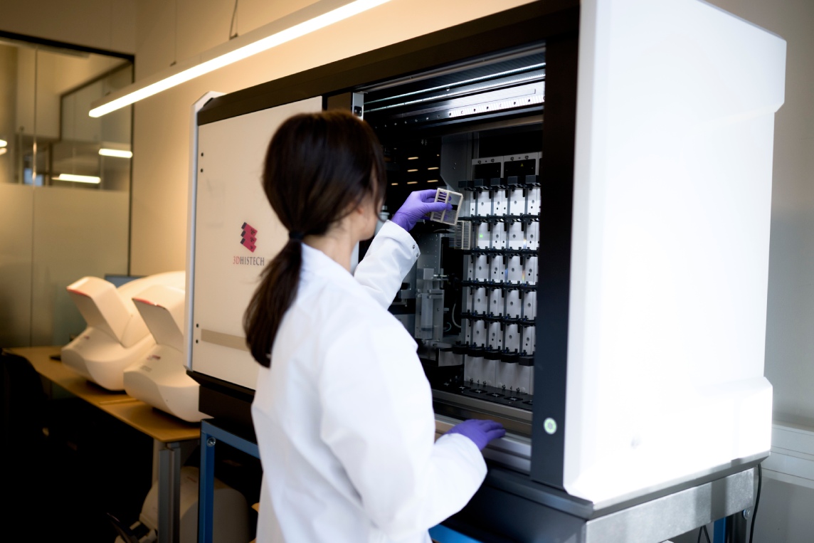
Access high-throughput, high-resolution imaging with fast turnaround times using industry-standard brightfield and fluorescent scanners from:
Whole slide scanning is available as a standalone digitization service or as part of an end-to-end digital pathology workflow.
Capture valuable data from tissues, such as cell phenotypes, densities, and spatial biology, using a broad portfolio of validated image analysis platforms, including:
In addition to running commercially validated assays, our scientists develop and validate custom algorithms to extract the specific information your clinical program needs.
Achieve faster, more consistent tissue analysis with AI-powered image analysis, including for:
We can implement and validate 3rd party algorithms or deploy commercial algorithms for all phases of clinical trials. We can also develop and validate new algorithms if your biomarker program requires.
Benefit from a specialist pathology team with extensive experience in scoring a vast range of diagnostic assays in the context of clinical trials and biomarker testing.
Our pathologists receive regular training in both novel and approved scoring methods from CDx developers, while an internal quality program delivers skills sharing and scoring alignment across the global network.
CellCarta announces the launch of the Digital Pathology and AI Consortium, giving sponsors a single CRO-led, global AI-agnostic path to accelerate biomarker discovery, patient stratification, and clinical trial execution across therapeutic areas.







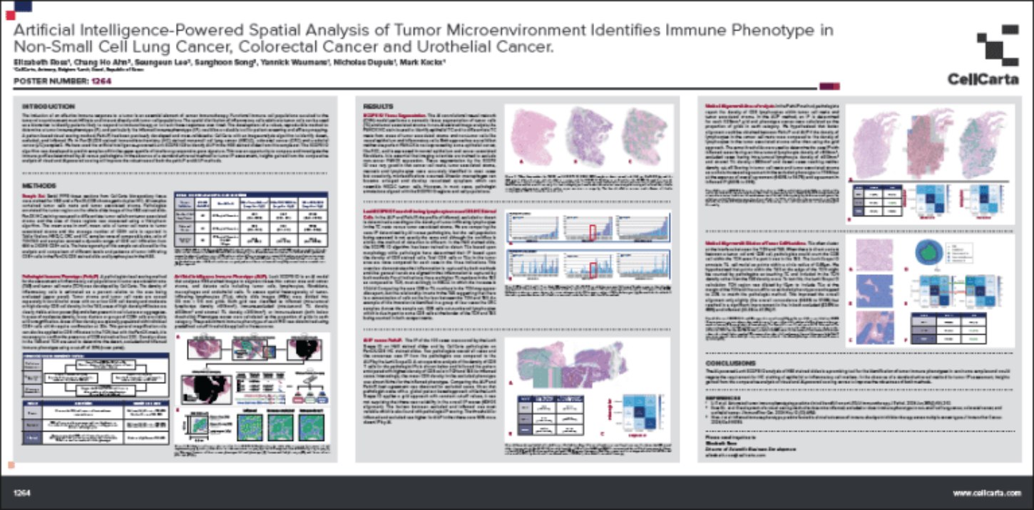
CellCarta performed a comparative analysis of the immune profiles of carcinoma samples when evaluated by the AI-powered Lunit SCOPE IO platform, pathologist visual assessment, and Visiopharm analysis. Insights gained here can improve the robustness of all methods for immune phenotype investigations.
Innovative digital pathology solutions to advance your clinical trial.
Platform-Agnostic Approach
20+ In-House Pathologists
Global Execution
AI-Powered Analysis
High-Touch Collaboration
Combine digital pathology solutions with complementary tissue-based services for a more complete picture of your biomarker data.
End-to-end histopathology services from a team with deep clinical trial expertise across multiple indications.
Reveal how cells interact in their native tissue environment with spatial biology tools built for clinical research.
Tissue biomarker data from 200+ validated IHC and IF assays scored by board-certified pathologists.
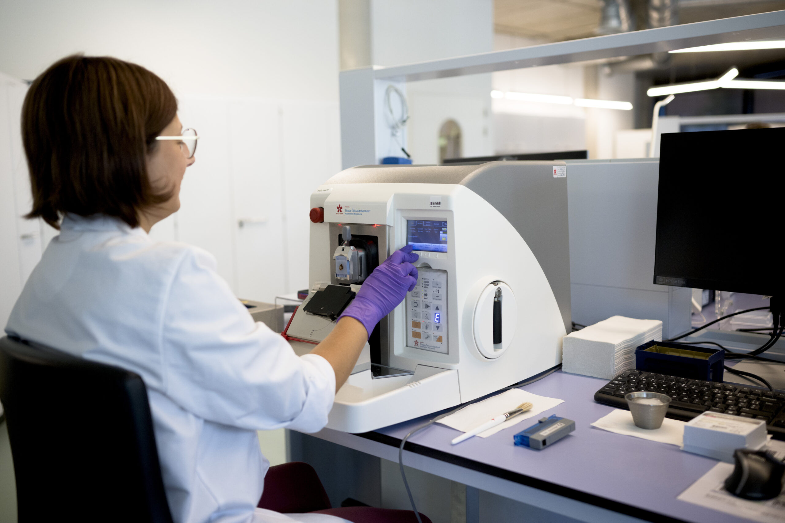

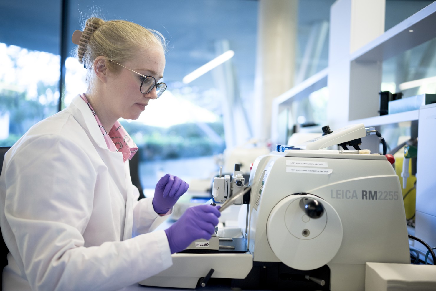
Our digital pathology solutions cover the full tissue analysis workflow, including whole slide brightfield and fluorescent scanning, multispectral imaging, multiplex immunofluorescence, and advanced image analysis. Services are available as standalone digitization or as part of an end-to-end workflow, all performed in a CLIA-certified, CAP-accredited laboratory by a dedicated team of pathologists and imaging scientists.
Digital pathology solutions can also be combined with broader histopathology and spatial biology services for a more complete tissue-based biomarker strategy.
CellCarta uses industry-standard whole slide scanning technologies from 3DHistech, Akoya, BioView, Leica Biosystems (Aperio®), Roche/Ventana, and Zeiss, enabling high-throughput, high-resolution brightfield and fluorescent imaging with fast turnaround times. Whole slide scanning is available as a standalone service for digitization programs or as part of an end-to-end digital pathology workflow.
Consistency is maintained through standardized SOPs, a rigorous internal pathology quality program, and the Pathotrainer platform, which supports scoring alignment, annotation, and continuing education across the global pathology network. Pathologists receive regular training in both novel and approved scoring methods from CDx developers, ensuring aligned scoring regardless of site.
AI is embedded across all of CellCarta's image analysis platforms, supporting nuclear segmentation, tissue classification, cell phenotyping, and circulating tumor cell detection. Our team offers expertise in developing and validating new AI-based algorithms for study-specific endpoints, and recently completed a global deployment of HALO AI to further advance quantitative image analysis capabilities across our digital pathology services.
Yes. CellCarta's imaging scientists have expertise in developing and validating custom image analysis algorithms tailored to specific study endpoints. Our digital pathology platforms include HALO®, Visiopharm®, Lunit SCOPE®, inForm®, CyteHub®, and navify® by Roche, and our team can build bespoke algorithms or collaborate with your preferred digital pathology AI provider to apply their algorithms in clinical trial assays conducted at CellCarta.