August 13, 2024
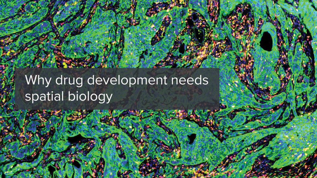
Understanding how drug candidates interact within diverse cellular environments and how these interactions translate to patient outcomes can be challenging. Moreover, heterogeneity within seemingly uniform diseases can lead to varied treatment responses, complicating the path to truly personalized therapies. As the field grapples with these challenges, new methodologies are needed to unravel the intricacies of disease and drug response. To this end, attention is increasingly turning to spatial biology.
Spatial biology is the study of cells within their native environments. Unlike traditional bulk approaches, spatial biology preserves the spatial context, allowing scientists to study how cells and molecules are distributed and interact within complex tissues and the architectural changes associated with various biological processes and diseases. By leveraging these insights, spatial biology enhances the understanding of drug effects at the cellular level, aiding the development of targeted therapies.
While spatial biology is not a new concept recent advances in multiplexing have transformed the field. These innovations enable the simultaneous measurement of multiple markers, and with enhanced computational capabilities, we can more effectively analyze the relationships between them. This deeper understanding of complex biological paradigms has elevated spatial biology into a powerful tool for drug development.
Drug developers are turning to spatial biology because it offers a deeper understanding of cellular context, leading to more accurate drug targeting, better predictions of drug efficacy, and insights into disease mechanisms.
As drug development is complex and costly, these comprehensive tools are essential for improving the efficiency of therapy development. Integrating spatial biology into drug development workflows offers several benefits:
Spatial biology has emerged as a groundbreaking discovery toolbox, significantly accelerating drug development by offering detailed insights into tissue microenvironments and molecular interactions. Cutting-edge platforms like Visium and Visium HD from 10X Genomics, GeoMX Digital Spatial Profiler from NanoString, and COMET from Lunaphore are at the forefront of this revolution. These platforms allow researchers to simultaneously capture high-plex RNA and protein information from a single tissue section, enabling a comprehensive understanding of cellular contexts and interactions.
Visium HD delivers high-resolution spatially resolved gene expression profiles, while GeoMX offers high-plex protein and RNA analysis powered by precise segmentation capabilities. COMET complements these with its sub-cellular resolution imaging capabilities and allows for the detailed exploration of tissue architecture and biomolecular interactions.
These tools are crucial for early clinical research as they rapidly generate valuable insights into disease mechanisms, drug efficacy, and biomarker validation.
By facilitating the exploration of complex tissue environments and enabling a deeper understanding of cellular heterogeneity, spatial biology tools streamline the path from preclinical studies to clinical application. This accelerates the identification of promising therapeutic candidates and optimizes their development, ensuring that drug developers can address key challenges more efficiently. With the path to clinical implementation becoming increasingly accessible, these advanced spatial biology platforms are poised to play a pivotal role in shaping the future of personalized medicine and therapeutic innovation.
To learn more about spatial biology and how it could help support and accelerate your therapeutic development, visit: spatial biology
About the author

İlayda Hasakioğulları is a Senior R&D Product Manager at CellCarta. Ilayda is passionate about precision medicine and specializes in spatial biology technologies. She continually scans the landscape to implement cutting-edge solutions at CellCarta and assists scientists in selecting the best tools for their research needs with the ultimate goal of benefiting patients.
August 13, 2024
Understanding how drug candidates interact within diverse cellular environments and how these interactions translate to patient outcomes can be challenging. Moreover, heterogeneity within seemingly uniform diseases can lead to varied treatment responses, complicating the path to truly personalized therapies. As the field grapples with these challenges, new methodologies are needed to unravel the intricacies of disease and drug response. To this end, attention is increasingly turning to spatial biology.
Spatial biology is the study of cells within their native environments. Unlike traditional bulk approaches, spatial biology preserves the spatial context, allowing scientists to study how cells and molecules are distributed and interact within complex tissues and the architectural changes associated with various biological processes and diseases. By leveraging these insights, spatial biology enhances the understanding of drug effects at the cellular level, aiding the development of targeted therapies.
While spatial biology is not a new concept recent advances in multiplexing have transformed the field. These innovations enable the simultaneous measurement of multiple markers, and with enhanced computational capabilities, we can more effectively analyze the relationships between them. This deeper understanding of complex biological paradigms has elevated spatial biology into a powerful tool for drug development.
Drug developers are turning to spatial biology because it offers a deeper understanding of cellular context, leading to more accurate drug targeting, better predictions of drug efficacy, and insights into disease mechanisms.
As drug development is complex and costly, these comprehensive tools are essential for improving the efficiency of therapy development. Integrating spatial biology into drug development workflows offers several benefits:
Spatial biology has emerged as a groundbreaking discovery toolbox, significantly accelerating drug development by offering detailed insights into tissue microenvironments and molecular interactions. Cutting-edge platforms like Visium and Visium HD from 10X Genomics, GeoMX Digital Spatial Profiler from NanoString, and COMET from Lunaphore are at the forefront of this revolution. These platforms allow researchers to simultaneously capture high-plex RNA and protein information from a single tissue section, enabling a comprehensive understanding of cellular contexts and interactions.
Visium HD delivers high-resolution spatially resolved gene expression profiles, while GeoMX offers high-plex protein and RNA analysis powered by precise segmentation capabilities. COMET complements these with its sub-cellular resolution imaging capabilities and allows for the detailed exploration of tissue architecture and biomolecular interactions.
These tools are crucial for early clinical research as they rapidly generate valuable insights into disease mechanisms, drug efficacy, and biomarker validation.
By facilitating the exploration of complex tissue environments and enabling a deeper understanding of cellular heterogeneity, spatial biology tools streamline the path from preclinical studies to clinical application. This accelerates the identification of promising therapeutic candidates and optimizes their development, ensuring that drug developers can address key challenges more efficiently. With the path to clinical implementation becoming increasingly accessible, these advanced spatial biology platforms are poised to play a pivotal role in shaping the future of personalized medicine and therapeutic innovation.
To learn more about spatial biology and how it could help support and accelerate your therapeutic development, visit: spatial biology
About the author
İlayda Hasakioğulları is a Senior R&D Product Manager at CellCarta. Ilayda is passionate about precision medicine and specializes in spatial biology technologies. She continually scans the landscape to implement cutting-edge solutions at CellCarta and assists scientists in selecting the best tools for their research needs with the ultimate goal of benefiting patients.
May 28, 2024
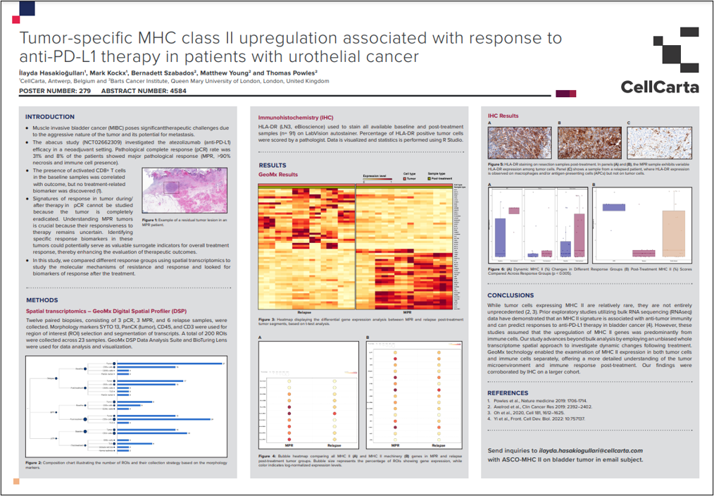
CellCarta’s poster, presented at ASCO 2024, discusses the study on tumor-specific MHC class II upregulation in patients with urothelial cancer undergoing anti-PD-L1 therapy.
The study, part of the abacus trial (NCT02662309), revealed that MHC class II expression in tumor cells, although rare, is associated with a positive response to atezolizumab. Utilizing spatial transcriptomics and immunohistochemistry (IHC), the research highlighted significant differences in MHC II expression between patients with pathological complete response (pCR) and those with major pathological response (MPR) or relapse.
The findings suggest that MHC II expression could serve as a biomarker for predicting therapeutic outcomes in bladder cancer treatment.
View the full poster:
May 28, 2024
CellCarta’s poster, presented at ASCO 2024, discusses the study on tumor-specific MHC class II upregulation in patients with urothelial cancer undergoing anti-PD-L1 therapy.
The study, part of the abacus trial (NCT02662309), revealed that MHC class II expression in tumor cells, although rare, is associated with a positive response to atezolizumab. Utilizing spatial transcriptomics and immunohistochemistry (IHC), the research highlighted significant differences in MHC II expression between patients with pathological complete response (pCR) and those with major pathological response (MPR) or relapse.
The findings suggest that MHC II expression could serve as a biomarker for predicting therapeutic outcomes in bladder cancer treatment.
View the full poster:
December 19, 2023
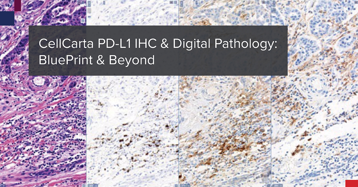
In October 2017, The BluePrint Phase 2 (BP2) committee presented to the International Association for the Study of Lung Cancer (IASLC) 18th World Conference on Lung Cancer. Together, they presented the data from the comparison of five PD-L1 immunohistochemistry (IHC) assays, that each utilize a different PD-L1 antibody clone: 22C3, 28-8, SP142, SP263, and 73-10. The data was published in the Journal of Thoracic Oncology.1 The goal was to validate the BP1 results using real-world clinical lung cancer samples.
The IHC staining for this BP2 comparison was performed in CellCarta’s CAP-accredited laboratory in Belgium. The company was then known as HistoGeneX. Whole slide images (WSIs) were prepared for the study and the scans were uploaded to PathoTrainer™.
Characteristics of PD-L1 Assays: Staining Performance
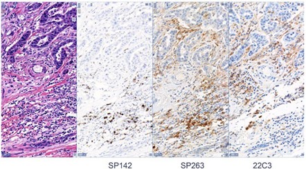
This software allowed for on-site and remote pathologist training in PD-L1 scoring, since the study also aimed to assess WSI analysis by a pathologist versus traditional light microscopic analysis. Mark Kockx, MD, PhD, a pathologist and founder of CellCarta HistoGeneX, participated in the study as a trainer and in slide scoring. Ultimately, the study confirmed the reliability of PD-L1 scoring of digital images. Since BP2 was published, the number of PD-1/L1 monoclonal antibody clinical trials has exploded.
The Cancer Research Institute created a dashboard of active interventional trials, with PD-1/-L1 agents and targets used in combo therapy trials. The graphs paint a colorful picture of the clinical trial landscape back in 2017 and again in 2021. In every category the number of clinical trials has increased. Pembrolizumab trials grew from 557 in 2017 to 1,481 in 2021, and the “Other PDx” category grew from 90 trials in 2017 to 1,631 in 2021.2 CRI counted 5,683 interventional trials as of December 2021.
Let’s fast forward now to the end of 2023.
CellCarta is performing PD-L1 IHC in Phase III trials more than any other biomarker, with all of the commercially available clones. PathoTrainer™ is still regularly used at CellCarta for in person and remote pathologist training, since it is an ideal tool for collaboration and for pathologist proficiency testing.
Scoring of PD-L1 is performed by trained pathologists for all established scoring algorithms including tumor proportion score (TPS), combined positive score (CPS), and tumor-infiltrating immune cell (IC) staining assessments. The turn- around time for this testing is as short as 3 business days.
As of 2022, CellCarta participated in more than 40 companion diagnostic (CDx) studies totaling over 70,000 slides. These numbers continue to increase.
There have also been many changes and advances in the field of digital pathology since BP2 was published. The outbreak of SARS-CoV-2 created a public health emergency that accelerated the need for policies to help reduce exposure of healthcare personnel. One way to do this was to expand the availability of remote digital pathology devices. CAP updated the original 2013 WSI guidelines in 2021 and these were published in The Archives of Pathology & Laboratory Medicine.3
The 2023 IASCL World Conference on Lung Cancer was in Singapore in September. Dr. Ming-Sound Tsao who is the globally recognized leader in molecular testing in lung cancer, and the lead author of the BP2 study, presented.
CellCarta has grown and evolved since BP2. Our compliant digital pathology-based workflows are now in use in our newer labs in the United States and China. Our global team of pathologists has grown to nearly 30. More companies are testing combination regimens like Antibody Drug Conjugates with Immunotherapy and Chemotherapy. As this field of drug development evolves, CellCarta will continue to provide leadership, and participate in the industry’s most significant targeted and immunotherapy biomarker programs.
About the author:

Sharron Webster, BS, MS, HTL(ASCP) is a Scientific Business Director for the Histopathology Services unit within CellCarta. As an ASCP certified histotechnologist, she has 20+ years of experience in histopathology laboratories, with a focus on immunohistochemistry development and validation. At CellCarta, Shar is using her expertise to help our clients find the best scientific and technical solutions for FFPE tissue and CTC analyses.
December 19, 2023
In October 2017, The BluePrint Phase 2 (BP2) committee presented to the International Association for the Study of Lung Cancer (IASLC) 18th World Conference on Lung Cancer. Together, they presented the data from the comparison of five PD-L1 immunohistochemistry (IHC) assays, that each utilize a different PD-L1 antibody clone: 22C3, 28-8, SP142, SP263, and 73-10. The data was published in the Journal of Thoracic Oncology.1 The goal was to validate the BP1 results using real-world clinical lung cancer samples.
The IHC staining for this BP2 comparison was performed in CellCarta’s CAP-accredited laboratory in Belgium. The company was then known as HistoGeneX. Whole slide images (WSIs) were prepared for the study and the scans were uploaded to PathoTrainer™.
Characteristics of PD-L1 Assays: Staining Performance
This software allowed for on-site and remote pathologist training in PD-L1 scoring, since the study also aimed to assess WSI analysis by a pathologist versus traditional light microscopic analysis. Mark Kockx, MD, PhD, a pathologist and founder of CellCarta HistoGeneX, participated in the study as a trainer and in slide scoring. Ultimately, the study confirmed the reliability of PD-L1 scoring of digital images. Since BP2 was published, the number of PD-1/L1 monoclonal antibody clinical trials has exploded.
The Cancer Research Institute created a dashboard of active interventional trials, with PD-1/-L1 agents and targets used in combo therapy trials. The graphs paint a colorful picture of the clinical trial landscape back in 2017 and again in 2021. In every category the number of clinical trials has increased. Pembrolizumab trials grew from 557 in 2017 to 1,481 in 2021, and the “Other PDx” category grew from 90 trials in 2017 to 1,631 in 2021.2 CRI counted 5,683 interventional trials as of December 2021.
Let’s fast forward now to the end of 2023.
CellCarta is performing PD-L1 IHC in Phase III trials more than any other biomarker, with all of the commercially available clones. PathoTrainer™ is still regularly used at CellCarta for in person and remote pathologist training, since it is an ideal tool for collaboration and for pathologist proficiency testing.
Scoring of PD-L1 is performed by trained pathologists for all established scoring algorithms including tumor proportion score (TPS), combined positive score (CPS), and tumor-infiltrating immune cell (IC) staining assessments. The turn- around time for this testing is as short as 3 business days.
As of 2022, CellCarta participated in more than 40 companion diagnostic (CDx) studies totaling over 70,000 slides. These numbers continue to increase.
There have also been many changes and advances in the field of digital pathology since BP2 was published. The outbreak of SARS-CoV-2 created a public health emergency that accelerated the need for policies to help reduce exposure of healthcare personnel. One way to do this was to expand the availability of remote digital pathology devices. CAP updated the original 2013 WSI guidelines in 2021 and these were published in The Archives of Pathology & Laboratory Medicine.3
The 2023 IASCL World Conference on Lung Cancer was in Singapore in September. Dr. Ming-Sound Tsao who is the globally recognized leader in molecular testing in lung cancer, and the lead author of the BP2 study, presented.
CellCarta has grown and evolved since BP2. Our compliant digital pathology-based workflows are now in use in our newer labs in the United States and China. Our global team of pathologists has grown to nearly 30. More companies are testing combination regimens like Antibody Drug Conjugates with Immunotherapy and Chemotherapy. As this field of drug development evolves, CellCarta will continue to provide leadership, and participate in the industry’s most significant targeted and immunotherapy biomarker programs.
About the author:
Sharron Webster, BS, MS, HTL(ASCP) is a Scientific Business Director for the Histopathology Services unit within CellCarta. As an ASCP certified histotechnologist, she has 20+ years of experience in histopathology laboratories, with a focus on immunohistochemistry development and validation. At CellCarta, Shar is using her expertise to help our clients find the best scientific and technical solutions for FFPE tissue and CTC analyses.
October 24, 2023
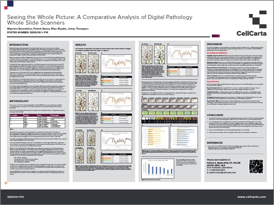
CellCarta’s poster compares the performance of different digital pathology whole slide scanners (WSI) to improve standardization in digital pathology.
The study evaluates the image quality, color accuracy, and reliability of various scanners using a standardized set of pathological samples. It highlights the strengths and weaknesses of each scanner, providing guidance for pathologists and researchers in selecting the most appropriate WSI format for their needs. The inclusion of AI-driven algorithms for marker detection, such as PD-L1, demonstrates the potential for automation and improved diagnostic accuracy in digital pathology.
For more insights on advanced digital pathology solutions and scanner contact us today
View the full poster:
Seeing the Whole Picture: A Comparative Analysis of Digital Pathology Whole Slide Scanners
October 24, 2023
CellCarta’s poster compares the performance of different digital pathology whole slide scanners (WSI) to improve standardization in digital pathology.
The study evaluates the image quality, color accuracy, and reliability of various scanners using a standardized set of pathological samples. It highlights the strengths and weaknesses of each scanner, providing guidance for pathologists and researchers in selecting the most appropriate WSI format for their needs. The inclusion of AI-driven algorithms for marker detection, such as PD-L1, demonstrates the potential for automation and improved diagnostic accuracy in digital pathology.
For more insights on advanced digital pathology solutions and scanner contact us today
View the full poster:
Seeing the Whole Picture: A Comparative Analysis of Digital Pathology Whole Slide Scanners