PD-L1 IHC and Digital Pathology: Insights and Future Directions

December 19, 2023
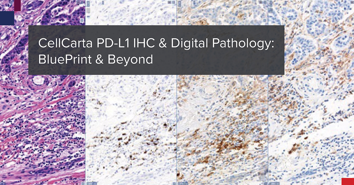
PD-L1 IHC & Digital Pathology: BluePrint & Beyond

HOW IT STARTED

In October 2017, The BluePrint Phase 2 (BP2) committee presented to the International Association for the Study of Lung Cancer (IASLC) 18th World Conference on Lung Cancer. Together, they presented the data from the comparison of five PD-L1 immunohistochemistry (IHC) assays, that each utilize a different PD-L1 antibody clone: 22C3, 28-8, SP142, SP263, and 73-10. The data was published in the Journal of Thoracic Oncology.1 The goal was to validate the BP1 results using real-world clinical lung cancer samples.

The IHC staining for this BP2 comparison was performed in CellCarta’s CAP-accredited laboratory in Belgium. The company was then known as HistoGeneX. Whole slide images (WSIs) were prepared for the study and the scans were uploaded to PathoTrainer™.

Characteristics of PD-L1 Assays: Staining Performance
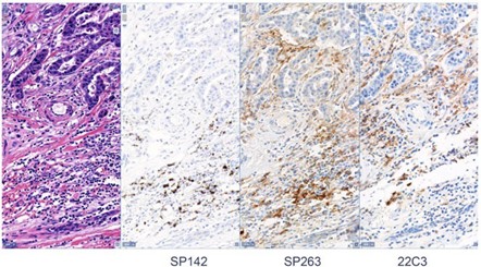
IHC PD-L1 stainings

This software allowed for on-site and remote pathologist training in PD-L1 scoring, since the study also aimed to assess WSI analysis by a pathologist versus traditional light  microscopic  analysis.  Mark Kockx, MD, PhD, a pathologist and founder of CellCarta HistoGeneX, participated in the study as a trainer and in slide scoring. Ultimately, the study confirmed the reliability of PD-L1 scoring of digital images. Since BP2 was published, the number of PD-1/L1 monoclonal antibody clinical trials has exploded.

The Cancer Research Institute created a dashboard of active interventional trials, with PD-1/-L1 agents and targets used in combo therapy trials. The graphs paint a colorful picture of the clinical trial landscape back in 2017 and again in 2021. In every category the number of clinical trials has increased. Pembrolizumab trials grew from 557 in 2017 to 1,481 in 2021, and the “Other PDx” category grew from 90 trials in 2017 to 1,631 in 2021.2 CRI counted 5,683 interventional trials as of December 2021.

HOW IT’S GOING

Let’s fast forward now to the end of 2023.

CellCarta is performing PD-L1 IHC in Phase III trials more than any other biomarker, with all of the commercially available clones. PathoTrainer™ is still regularly used at CellCarta for in person and remote pathologist training,  since it is an ideal tool for collaboration and for pathologist proficiency testing.

Scoring of PD-L1 is performed by trained pathologists for all established scoring algorithms including tumor proportion score (TPS), combined positive score (CPS), and tumor-infiltrating immune cell (IC) staining assessments. The turn- around time for this testing is as short as 3 business days.

As of 2022, CellCarta participated in more than 40 companion diagnostic (CDx) studies totaling over 70,000 slides. These numbers continue to increase.

There have also been many changes and advances in the field of digital pathology since BP2 was published. The outbreak of SARS-CoV-2 created a public health emergency that accelerated the need for policies to help reduce exposure of healthcare personnel. One way to do  this was to expand the availability of remote digital pathology devices. CAP updated the original 2013 WSI guidelines in 2021 and these were published in The Archives of Pathology & Laboratory Medicine.3

The 2023 IASCL World Conference on Lung Cancer was in Singapore in September. Dr. Ming-Sound Tsao who is the globally recognized leader in molecular testing in lung cancer, and the lead author of the BP2 study, presented.

WHAT’S NEXT?

CellCarta has grown and evolved since BP2. Our compliant digital pathology-based workflows are now in use in our newer labs in the United States and China. Our  global team of pathologists has grown to nearly 30. More companies are testing combination regimens like Antibody Drug Conjugates with Immunotherapy and Chemotherapy. As this field of drug development evolves, CellCarta will continue to provide leadership, and participate in the industry’s most significant targeted and immunotherapy biomarker programs.


About the author:

author photo

Sharron Webster, BS, MS, HTL(ASCP) is a Scientific Business Director for the Histopathology Services unit within CellCarta. As an ASCP certified histotechnologist, she has 20+ years of experience in histopathology laboratories, with a focus on immunohistochemistry development and validation. At CellCarta, Shar is using her expertise to help our clients find the best scientific and technical solutions for FFPE tissue and CTC analyses.

References & Additional Reading:

  1. PD-L1 Immunohistochemistry Comparability Study in Real-Life
    Clinical Samples: Results of Blueprint Phase 2 Project (jto.org)
  2. 2019-09 PD-1/L1 trial landscape dashboard | Tableau Public
  3. Validating Whole Slide Imaging Systems for Diagnostic
    Purposes in Pathology | Archives of Pathology & Laboratory
    Medicine (allenpress.com)
PD-L1 IHC and Digital Pathology: Insights and Future Directions

December 19, 2023

PD-L1 IHC & Digital Pathology: BluePrint & Beyond

HOW IT STARTED

In October 2017, The BluePrint Phase 2 (BP2) committee presented to the International Association for the Study of Lung Cancer (IASLC) 18th World Conference on Lung Cancer. Together, they presented the data from the comparison of five PD-L1 immunohistochemistry (IHC) assays, that each utilize a different PD-L1 antibody clone: 22C3, 28-8, SP142, SP263, and 73-10. The data was published in the Journal of Thoracic Oncology.1 The goal was to validate the BP1 results using real-world clinical lung cancer samples.

The IHC staining for this BP2 comparison was performed in CellCarta’s CAP-accredited laboratory in Belgium. The company was then known as HistoGeneX. Whole slide images (WSIs) were prepared for the study and the scans were uploaded to PathoTrainer™.

Characteristics of PD-L1 Assays: Staining Performance

IHC PD-L1 stainings

This software allowed for on-site and remote pathologist training in PD-L1 scoring, since the study also aimed to assess WSI analysis by a pathologist versus traditional light  microscopic  analysis.  Mark Kockx, MD, PhD, a pathologist and founder of CellCarta HistoGeneX, participated in the study as a trainer and in slide scoring. Ultimately, the study confirmed the reliability of PD-L1 scoring of digital images. Since BP2 was published, the number of PD-1/L1 monoclonal antibody clinical trials has exploded.

The Cancer Research Institute created a dashboard of active interventional trials, with PD-1/-L1 agents and targets used in combo therapy trials. The graphs paint a colorful picture of the clinical trial landscape back in 2017 and again in 2021. In every category the number of clinical trials has increased. Pembrolizumab trials grew from 557 in 2017 to 1,481 in 2021, and the “Other PDx” category grew from 90 trials in 2017 to 1,631 in 2021.2 CRI counted 5,683 interventional trials as of December 2021.

HOW IT’S GOING

Let’s fast forward now to the end of 2023.

CellCarta is performing PD-L1 IHC in Phase III trials more than any other biomarker, with all of the commercially available clones. PathoTrainer™ is still regularly used at CellCarta for in person and remote pathologist training,  since it is an ideal tool for collaboration and for pathologist proficiency testing.

Scoring of PD-L1 is performed by trained pathologists for all established scoring algorithms including tumor proportion score (TPS), combined positive score (CPS), and tumor-infiltrating immune cell (IC) staining assessments. The turn- around time for this testing is as short as 3 business days.

As of 2022, CellCarta participated in more than 40 companion diagnostic (CDx) studies totaling over 70,000 slides. These numbers continue to increase.

There have also been many changes and advances in the field of digital pathology since BP2 was published. The outbreak of SARS-CoV-2 created a public health emergency that accelerated the need for policies to help reduce exposure of healthcare personnel. One way to do  this was to expand the availability of remote digital pathology devices. CAP updated the original 2013 WSI guidelines in 2021 and these were published in The Archives of Pathology & Laboratory Medicine.3

The 2023 IASCL World Conference on Lung Cancer was in Singapore in September. Dr. Ming-Sound Tsao who is the globally recognized leader in molecular testing in lung cancer, and the lead author of the BP2 study, presented.

WHAT’S NEXT?

CellCarta has grown and evolved since BP2. Our compliant digital pathology-based workflows are now in use in our newer labs in the United States and China. Our  global team of pathologists has grown to nearly 30. More companies are testing combination regimens like Antibody Drug Conjugates with Immunotherapy and Chemotherapy. As this field of drug development evolves, CellCarta will continue to provide leadership, and participate in the industry’s most significant targeted and immunotherapy biomarker programs.


About the author:

author photo

Sharron Webster, BS, MS, HTL(ASCP) is a Scientific Business Director for the Histopathology Services unit within CellCarta. As an ASCP certified histotechnologist, she has 20+ years of experience in histopathology laboratories, with a focus on immunohistochemistry development and validation. At CellCarta, Shar is using her expertise to help our clients find the best scientific and technical solutions for FFPE tissue and CTC analyses.

References & Additional Reading:

  1. PD-L1 Immunohistochemistry Comparability Study in Real-Life
    Clinical Samples: Results of Blueprint Phase 2 Project (jto.org)
  2. 2019-09 PD-1/L1 trial landscape dashboard | Tableau Public
  3. Validating Whole Slide Imaging Systems for Diagnostic
    Purposes in Pathology | Archives of Pathology & Laboratory
    Medicine (allenpress.com)

How HLA Typing Improves Immunotherapy Precision

December 6, 2023

How can HLA typing drive better immunotherapy development and selection

Immunotherapies hold enormous potential for cancer treatment, but understanding tumor–immune interactions is needed to successfully develop new therapies and identify suitable patients.

Human leucocyte antigen (HLA) typing gives deeper insights into the relationship between the immune system and cancer cells, helping scientists improve immunotherapy administration precision and develop new immunotherapeutic approaches. But what is it?

A deeper dive into HLA typing

HLAs are a combination of different single nucleotide polymorphisms (SNPs) across multiple exons. The HLA system helps the immune system distinguish between self and non-self cells by presenting antigens derived from pathogens, cancer cells, or other foreign entities to immune cells, triggering a response. HLAs are therefore pivotal in cancer immunotherapy — cancer-specific antigens are presented on HLA molecules, which T cells recognize, attack, and kill.

HLA allele and antigen composition varies significantly between individuals, however, so determining their HLA profile — HLA typing — is essential to understand disease onset, progression, and treatment.

How is HLA typing performed?

Most researchers use Sanger sequencing for HLA typing, which detects SNPs. But since both copies of the genes are sequenced together in one reaction, researchers struggle to identify whether the SNPs come from the same (cis) or the opposite (trans) chromosome, making establishing the correct HLA type difficult. Next-generation sequencing (NGS) overcomes cis/trans polymorphism ambiguities by independently identifying and amplifying individual DNA fragments for more accurate typing.

HLA typing in action

Three recent publications highlight the impact of HLA profiling in immunotherapy development:

1.    Immunotherapy in oncology

HLA type affects an individual’s susceptibility to infections or autoimmune diseases, or their oncological immunotherapy response. A 2019 publication discussed how understanding a patient’s HLA profile is vital to effectively tailor immunotherapeutic treatments. Particularly, researchers should match the HLA types of cancer patients with potential donor HLA types to improve the success of treatments like adoptive cell therapy or stem cell therapy.

HLA typing is essential in CAR T-cell therapy, where it helps in the engineering of T cells to target tumor antigens, expanding immunotherapy applications beyond standard treatments.

2.    Precision medicine

HLA genotyping can help researchers to predict the survival of HLA alleles among cancers, based on an underlying cytolytic activity (CYT) mechanism. A recent study showed that a strong HLA allele, in combination with a high tumor mutation burden, could stimulate intensive immune CYT and lead to extended HLA survival. As HLA profiles differ between patients, typing can therefore support oncologists in selecting the most appropriate immunotherapy for an individual.

3.    Viral associated cancer

The 2019 article also highlighted that carcinogenic viruses activate genes that allow cancer cells to escape immune surveillance and proliferate uncontrolled. A patient’s HLA profile plays a significant role in these immune escape mechanisms, leading to the initiation of such cancers and influencing immunotherapy response. Understanding HLA profiles can therefore help effective cancer vaccine design, improving patient outcomes.

Immunotherapy is a promising approach to treat cancers — and HLA typing is key to unlocking its potential. With HLA typing, researchers can better tailor therapies, select the right treatment, and design more effective cancer vaccines.

NGS is critical to getting clearer results from your HLA typing.

Future of HLA Typing: Artificial Intelligence and Predictive Algorithms

As HLA typing evolves, the integration of AI and machine learning is becoming key to advancing immunotherapy.

These technologies are being used to develop predictive models that combine HLA typing with tumor mutational burden (TMB), and other biomarkers, to forecast patient responses to immune checkpoint inhibitors.

This multidisciplinary approach is expected to revolutionize the personalization of cancer treatments.

At CellCarta we developed our own data analysis pipeline to evaluate immune checkpoint biomarkers by looking at RNA sequencing data.

Discover CellCarta’s genomics services.


About the author: 

Author photo

Nathalie Bernard (PhD) is the scientific business director for the Genomics Services unit within CellCarta. Her background is in molecular biology, and she has many years of experience in PCR and sequencing, technologies used to discover or identify DNA and RNA biomarkers of clinical utility. At CellCarta, Nathalie is using her expertise to guide our customers in finding the best solution to their genomic questions.

How HLA Typing Improves Immunotherapy Precision

December 6, 2023

How can HLA typing drive better immunotherapy development and selection

Immunotherapies hold enormous potential for cancer treatment, but understanding tumor–immune interactions is needed to successfully develop new therapies and identify suitable patients.

Human leucocyte antigen (HLA) typing gives deeper insights into the relationship between the immune system and cancer cells, helping scientists improve immunotherapy administration precision and develop new immunotherapeutic approaches. But what is it?

A deeper dive into HLA typing

HLAs are a combination of different single nucleotide polymorphisms (SNPs) across multiple exons. The HLA system helps the immune system distinguish between self and non-self cells by presenting antigens derived from pathogens, cancer cells, or other foreign entities to immune cells, triggering a response. HLAs are therefore pivotal in cancer immunotherapy — cancer-specific antigens are presented on HLA molecules, which T cells recognize, attack, and kill.

HLA allele and antigen composition varies significantly between individuals, however, so determining their HLA profile — HLA typing — is essential to understand disease onset, progression, and treatment.

How is HLA typing performed?

Most researchers use Sanger sequencing for HLA typing, which detects SNPs. But since both copies of the genes are sequenced together in one reaction, researchers struggle to identify whether the SNPs come from the same (cis) or the opposite (trans) chromosome, making establishing the correct HLA type difficult. Next-generation sequencing (NGS) overcomes cis/trans polymorphism ambiguities by independently identifying and amplifying individual DNA fragments for more accurate typing.

HLA typing in action

Three recent publications highlight the impact of HLA profiling in immunotherapy development:

1.    Immunotherapy in oncology

HLA type affects an individual’s susceptibility to infections or autoimmune diseases, or their oncological immunotherapy response. A 2019 publication discussed how understanding a patient’s HLA profile is vital to effectively tailor immunotherapeutic treatments. Particularly, researchers should match the HLA types of cancer patients with potential donor HLA types to improve the success of treatments like adoptive cell therapy or stem cell therapy.

HLA typing is essential in CAR T-cell therapy, where it helps in the engineering of T cells to target tumor antigens, expanding immunotherapy applications beyond standard treatments.

2.    Precision medicine

HLA genotyping can help researchers to predict the survival of HLA alleles among cancers, based on an underlying cytolytic activity (CYT) mechanism. A recent study showed that a strong HLA allele, in combination with a high tumor mutation burden, could stimulate intensive immune CYT and lead to extended HLA survival. As HLA profiles differ between patients, typing can therefore support oncologists in selecting the most appropriate immunotherapy for an individual.

3.    Viral associated cancer

The 2019 article also highlighted that carcinogenic viruses activate genes that allow cancer cells to escape immune surveillance and proliferate uncontrolled. A patient’s HLA profile plays a significant role in these immune escape mechanisms, leading to the initiation of such cancers and influencing immunotherapy response. Understanding HLA profiles can therefore help effective cancer vaccine design, improving patient outcomes.

Immunotherapy is a promising approach to treat cancers — and HLA typing is key to unlocking its potential. With HLA typing, researchers can better tailor therapies, select the right treatment, and design more effective cancer vaccines.

NGS is critical to getting clearer results from your HLA typing.

Future of HLA Typing: Artificial Intelligence and Predictive Algorithms

As HLA typing evolves, the integration of AI and machine learning is becoming key to advancing immunotherapy.

These technologies are being used to develop predictive models that combine HLA typing with tumor mutational burden (TMB), and other biomarkers, to forecast patient responses to immune checkpoint inhibitors.

This multidisciplinary approach is expected to revolutionize the personalization of cancer treatments.

At CellCarta we developed our own data analysis pipeline to evaluate immune checkpoint biomarkers by looking at RNA sequencing data.

Discover CellCarta’s genomics services.


About the author: 

Author photo

Nathalie Bernard (PhD) is the scientific business director for the Genomics Services unit within CellCarta. Her background is in molecular biology, and she has many years of experience in PCR and sequencing, technologies used to discover or identify DNA and RNA biomarkers of clinical utility. At CellCarta, Nathalie is using her expertise to guide our customers in finding the best solution to their genomic questions.

Enhance Flow Cytometry with Automatic Gating Tools

November 15, 2023

Automatic Gating Tools- autogating, range gating

Gating can be one of the more time-consuming parts of cytometry analysis. When gate positions need to be changed for every sample in an experiment, it can take orders of magnitude longer.

Two automatic tools built into CellEngine, CellCarta’s cytometry analysis platform, can be leveraged to increase speed and efficiency:

  • Automatic gate tailoring uses a machine learning algorithm to adjust gate placement. After defining a population hierarchy, select one or more gates to be adjusted to fit each sample’s data automatically. A set of manually gated reference files provide a model for the algorithm to position gates in the rest of the data. When faced with datasets with high variability, automatic gate tailoring is one of the most powerful tools to expedite analysis.
  • Automatic percentile gating precisely moves a gate vertex or edge to a specified percentile of one or more control files. For example, you can move a range gate to the 99th percentile of an unstimulated or isotype control. This approach facilitates comparative analysis of samples; for example, when comparing normalized outcomes donor-by-donor.

In order to streamline the gating process most efficiently, there are several important points to remember when using these tools.

1 – Understanding Automatic Gating Tools in Flow Cytometry with CellEngine

Experimental design is a fundamental factor for successful cytometry analysis. Autogating in CellEngine streamlines the analysis process, but the best results come from well-designed trials and high-quality data.

Taking time to design clear objectives, include appropriate controls, and optimize experimental conditions ensures that the results from autogating are meaningful and useful.

For example, including an unstimulated control in an intracellular cytokine or phospho-flow panel provides an ideal control for using percentile gating.

Picking dyes that provide good separation of populations improves automatic gate tailoring’s performance. By creating a well-designed foundation, researchers can extract meaningful insights from complex datasets.

2 – Quality control is indispensable for Flow Cytometry Analysis

One of the most important aspects of high-quality analysis is high-quality data. To generate high-quality data, build quality control processes into your workflows to ensure reliability and accuracy, and identify issues promptly.

For example, verify staining via positive controls, use beads to calibrate and verify cytometer performance, and reduce technical variability as much as possible.

This maximizes the potential from each dataset, whether you use automatic tools in analysis or not.

3 – How to Implement Automatic Gating in CellEngine

Automatic gate tailoring uses one or more files as a reference for setting gate positions. Training data must be representative of the overall dataset, and carefully gated for best results.

An experiment with different cell types, disease states, or experimental conditions should include examples of all of those in the training data. This enhances the algorithm’s ability to properly adjust gates.

Consistent choices when gating the training data will also reduce variability.

Combining meticulous experiment design, trustworthy data, and carefully curated training data with automated tools increases efficiency and accuracy.

By incorporating these best practices, researchers can harness the full power of autogating tools to optimize their workflows.

If you’re interested in trying autogating, you can sign up for a free two-month trial of CellEngine at https://cellengine.com/.


About the author:

author photo

Anita Ray is a technical application specialist with CellEngine. She has ten years of experience in cytometry and worked in translational immuno-oncology prior to joining the CellEngine team.

Enhance Flow Cytometry with Automatic Gating Tools

November 15, 2023

Automatic Gating Tools- autogating, range gating

Gating can be one of the more time-consuming parts of cytometry analysis. When gate positions need to be changed for every sample in an experiment, it can take orders of magnitude longer.

Two automatic tools built into CellEngine, CellCarta’s cytometry analysis platform, can be leveraged to increase speed and efficiency:

  • Automatic gate tailoring uses a machine learning algorithm to adjust gate placement. After defining a population hierarchy, select one or more gates to be adjusted to fit each sample’s data automatically. A set of manually gated reference files provide a model for the algorithm to position gates in the rest of the data. When faced with datasets with high variability, automatic gate tailoring is one of the most powerful tools to expedite analysis.
  • Automatic percentile gating precisely moves a gate vertex or edge to a specified percentile of one or more control files. For example, you can move a range gate to the 99th percentile of an unstimulated or isotype control. This approach facilitates comparative analysis of samples; for example, when comparing normalized outcomes donor-by-donor.

In order to streamline the gating process most efficiently, there are several important points to remember when using these tools.

1 – Understanding Automatic Gating Tools in Flow Cytometry with CellEngine

Experimental design is a fundamental factor for successful cytometry analysis. Autogating in CellEngine streamlines the analysis process, but the best results come from well-designed trials and high-quality data.

Taking time to design clear objectives, include appropriate controls, and optimize experimental conditions ensures that the results from autogating are meaningful and useful.

For example, including an unstimulated control in an intracellular cytokine or phospho-flow panel provides an ideal control for using percentile gating.

Picking dyes that provide good separation of populations improves automatic gate tailoring’s performance. By creating a well-designed foundation, researchers can extract meaningful insights from complex datasets.

2 – Quality control is indispensable for Flow Cytometry Analysis

One of the most important aspects of high-quality analysis is high-quality data. To generate high-quality data, build quality control processes into your workflows to ensure reliability and accuracy, and identify issues promptly.

For example, verify staining via positive controls, use beads to calibrate and verify cytometer performance, and reduce technical variability as much as possible.

This maximizes the potential from each dataset, whether you use automatic tools in analysis or not.

3 – How to Implement Automatic Gating in CellEngine

Automatic gate tailoring uses one or more files as a reference for setting gate positions. Training data must be representative of the overall dataset, and carefully gated for best results.

An experiment with different cell types, disease states, or experimental conditions should include examples of all of those in the training data. This enhances the algorithm’s ability to properly adjust gates.

Consistent choices when gating the training data will also reduce variability.

Combining meticulous experiment design, trustworthy data, and carefully curated training data with automated tools increases efficiency and accuracy.

By incorporating these best practices, researchers can harness the full power of autogating tools to optimize their workflows.

If you’re interested in trying autogating, you can sign up for a free two-month trial of CellEngine at https://cellengine.com/.


About the author:

author photo

Anita Ray is a technical application specialist with CellEngine. She has ten years of experience in cytometry and worked in translational immuno-oncology prior to joining the CellEngine team.

Navigating IVDR: How It Impacts Your Clinical Trial Assays

June 28, 2023

IVDR

In Vitro Diagnostics (IVDs) are an essential part of healthcare, helping clinicians screen, diagnose, monitor, and treat various conditions. The In Vitro Diagnostic Regulation (IVDR) aims to improve the quality, safety, efficacy, and performance of IVD devices by increasing the requirements for their evaluation, regulation, and surveillance, thus significantly impacting clinical trials1-7.

The IVDR, introduced by the European Union (EU), came into effect in May 26, 2022 and replaced the previously established In Vitro Diagnostic Directive (IVDD)1,3. With increased regulatory oversight, the IVDR encompasses more stringent requirements for the designation of Notified Bodies (NBs) and monitoring by the national competent authorities and the Commission1–5. Thus, IVDR reduces the risks of discrepancies in interpretation across the EU market and enables timely detection of safety issues via post-market traceability and transparency. Unlike the IVDD, the IVDR places greater emphasis on product-life cycle management and continuous product evaluation. Manufacturers are obligated to demonstrate the implementation of an effective quality management system (QMS)5.

Successful IVD development requires a knowledgeable team. At CellCarta, we have the expertise to navigate the process of IVD development and provide quality and regulatory solutions for IVDR challenges.

How does IVDR impact clinical trial assays?

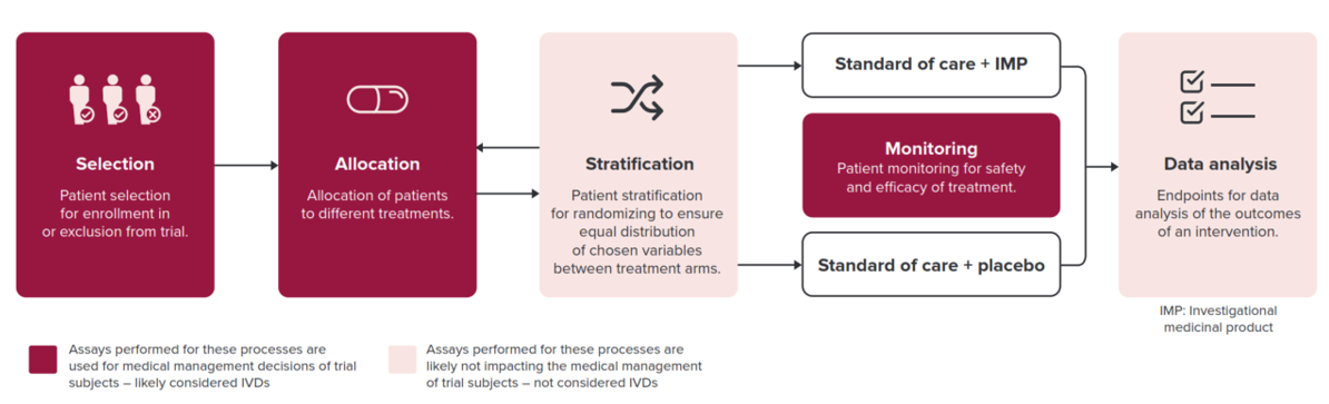
Within a clinical trial, all assays with a medical purpose are considered an IVD and are subject to IVDR3. More particularly, clinical trial assays used for medical management decisions of European subjects within the trial, involving patient selection, treatment allocation, and safety monitoring fall under IVD regulation (Figure 1). Thus, it is crucial to have a comprehensive understanding of the legislative requirements, and the devices affected, prior to initiating clinical trials. In-depth IVDR expertise minimizes the need for remediation following initial submission for performance evaluation and reduces the risk of commercial product withdrawal.

Section image

Figure 1. Processes determining which clinical assays are deemed IVDs and thus subject to IVDR. Exploratory assays for which correlation with clinical parameters is investigated retrospectively without having an impact on patients’ treatment are not subject to IVDR.

The shift from IVDD to IVDR brings about increased scrutiny of the performance evaluation plan concerning scientific validity, analytical performance, and clinical performance data5. The IVDR requires generation of clinical evidence to demonstrate conformity for all IVD devices, including those that were previously exempted under the IVDD3-5. As follows, clinical trial assays will need to provide more detailed and robust data on the performance of IVDs, including sensitivity, specificity, and predictive values2,4.

Additionally, IVDR introduces a new classification system for IVD devices, which considers the risks associated with the use of the device5. Some IVD devices previously classified as low-risk may now be classified as medium or high-risk and will require more extensive clinical trial data to obtain regulatory approval1,2,3,5.

The European Database on Medical Devices (EUDAMED) is a key aspect of rules established for IVDs under IVDR and is intended to improve transparency, traceability, and coordination among regulatory authorities and economic operations within the EU7. It enables efficient monitoring of devices throughout their lifecycle, enhances post-market surveillance, and strengthens patient safety.

What are Companion Diagnostics (CDx) devices?

Companion diagnostic (CDx) devices are a specific category of In Vitro Diagnostics that provide essential information for the safe and effective use of corresponding therapeutic products. CDx devices are categorized as Class C, representing a significant level of inherent risk6. At CellCarta, we specialize in providing comprehensive expertise in assay development and validation, guiding you through the transition of your assay from investigational clinical trial assay to a fully compliant IVD CDx.

Common questions, expert responses!

Is IVDR applicable for clinical trial testing in US-based laboratories?

  • IVDR is applicable whenever testing occurs on EU patient samples, irrespective of laboratory location. IVDR is applicable when a testing laboratory employs clinical trial assays developed in-house or utilizes a CE-IVD kit for off-label use, provided that the test results serve a medical purpose.

Do clinical trials initiated before May 26, 2022 require a performance study application under IVDR?

  • No, based on competent authority feedback, a performance study application is not required under IVDR if study subjects consented to participation in the trial before May 26, 2022.

Are all assays used in a clinical trial subject to IVD legislation?

  • No, exploratory assays not intended for medical management decisions of patient care or treatment, are not subject to IVDR. These include stratification and endpoint analysis, or other exploratory assays, for which correlation with clinical parameters is investigated retrospectively without impact on patient treatment (medical purpose).

CellCarta’s team of quality, regulatory and assay development experts understand the challenges of IVD CDx development and work with you to implement effective strategies.

Contact us for guidance through the complex IVDR landscape and to receive personalized support for your clinical trial projects.


About the author:

author photo

Sarah Berwouts is a Director Quality at the CellCarta Antwerp site, leading the ISO 13485 implementation for the development of Histopathology and Genomics assays. Sarah has over 20 years of professional experience, including 8 years in medical laboratory quality systems, and coordination of the European proficiency testing scheme for cystic fibrosis. She has an 8 additional years of expertise in the development and manufacturing of IVDs at Multiplicom and Agilent Technologies, in different roles in R&D and Quality.  Sarah has contributed to the implementation, maintenance, and harmonization of quality management systems under ISO 13485, ISO 15189, and ISO 17025. She studied Biomedical Sciences and received her PhD in quality improvement in medical laboratories.

References

  1. Lex – 32017R0746 – en – EUR-lex. EUR Available at: https://eur-lex.europa.eu/eli/reg/2017/746/oj. (Accessed: 26th June 2023)
  2. What’s changed compared to the IVDD – The European Union In Vitro Diagnostics Regulation. https://euivdr.com/whats-changed/.
  3. MDCG 2022-10 Q&A on the interface between Regulation (EU) 536/2014 on clinical trials for medicinal products for human use (CTR) and Regulation (EU) 2017/746 on in vitro diagnostic medical devices (IVDR). (2022).
  4. Dombrink, I. et al. Critical Implications of IVDR for Innovation in Diagnostics: Input From the BioMed Alliance Diagnostics Task Force. Hemasphere 6, E724 (2022).
  5. Factsheet for manufacturers of in vitro diagnostic. https://health.ec.europa.eu/system/files/2020-09/ivd_manufacturers_factsheet_en_0.pdf.
  6. Valla, V. et al. Companion Diagnostics: State of the Art and New Regulations. Biomark Insights 16, (2021).
  7. EUDAMED database – EUDAMED. https://ec.europa.eu/tools/eudamed/#/screen/home.

Navigating IVDR: How It Impacts Your Clinical Trial Assays

June 28, 2023

IVDR

In Vitro Diagnostics (IVDs) are an essential part of healthcare, helping clinicians screen, diagnose, monitor, and treat various conditions. The In Vitro Diagnostic Regulation (IVDR) aims to improve the quality, safety, efficacy, and performance of IVD devices by increasing the requirements for their evaluation, regulation, and surveillance, thus significantly impacting clinical trials1-7.

The IVDR, introduced by the European Union (EU), came into effect in May 26, 2022 and replaced the previously established In Vitro Diagnostic Directive (IVDD)1,3. With increased regulatory oversight, the IVDR encompasses more stringent requirements for the designation of Notified Bodies (NBs) and monitoring by the national competent authorities and the Commission1–5. Thus, IVDR reduces the risks of discrepancies in interpretation across the EU market and enables timely detection of safety issues via post-market traceability and transparency. Unlike the IVDD, the IVDR places greater emphasis on product-life cycle management and continuous product evaluation. Manufacturers are obligated to demonstrate the implementation of an effective quality management system (QMS)5.

Successful IVD development requires a knowledgeable team. At CellCarta, we have the expertise to navigate the process of IVD development and provide quality and regulatory solutions for IVDR challenges.

How does IVDR impact clinical trial assays?

Within a clinical trial, all assays with a medical purpose are considered an IVD and are subject to IVDR3. More particularly, clinical trial assays used for medical management decisions of European subjects within the trial, involving patient selection, treatment allocation, and safety monitoring fall under IVD regulation (Figure 1). Thus, it is crucial to have a comprehensive understanding of the legislative requirements, and the devices affected, prior to initiating clinical trials. In-depth IVDR expertise minimizes the need for remediation following initial submission for performance evaluation and reduces the risk of commercial product withdrawal.

Section image

Figure 1. Processes determining which clinical assays are deemed IVDs and thus subject to IVDR. Exploratory assays for which correlation with clinical parameters is investigated retrospectively without having an impact on patients’ treatment are not subject to IVDR.

The shift from IVDD to IVDR brings about increased scrutiny of the performance evaluation plan concerning scientific validity, analytical performance, and clinical performance data5. The IVDR requires generation of clinical evidence to demonstrate conformity for all IVD devices, including those that were previously exempted under the IVDD3-5. As follows, clinical trial assays will need to provide more detailed and robust data on the performance of IVDs, including sensitivity, specificity, and predictive values2,4.

Additionally, IVDR introduces a new classification system for IVD devices, which considers the risks associated with the use of the device5. Some IVD devices previously classified as low-risk may now be classified as medium or high-risk and will require more extensive clinical trial data to obtain regulatory approval1,2,3,5.

The European Database on Medical Devices (EUDAMED) is a key aspect of rules established for IVDs under IVDR and is intended to improve transparency, traceability, and coordination among regulatory authorities and economic operations within the EU7. It enables efficient monitoring of devices throughout their lifecycle, enhances post-market surveillance, and strengthens patient safety.

What are Companion Diagnostics (CDx) devices?

Companion diagnostic (CDx) devices are a specific category of In Vitro Diagnostics that provide essential information for the safe and effective use of corresponding therapeutic products. CDx devices are categorized as Class C, representing a significant level of inherent risk6. At CellCarta, we specialize in providing comprehensive expertise in assay development and validation, guiding you through the transition of your assay from investigational clinical trial assay to a fully compliant IVD CDx.

Common questions, expert responses!

Is IVDR applicable for clinical trial testing in US-based laboratories?

  • IVDR is applicable whenever testing occurs on EU patient samples, irrespective of laboratory location. IVDR is applicable when a testing laboratory employs clinical trial assays developed in-house or utilizes a CE-IVD kit for off-label use, provided that the test results serve a medical purpose.

Do clinical trials initiated before May 26, 2022 require a performance study application under IVDR?

  • No, based on competent authority feedback, a performance study application is not required under IVDR if study subjects consented to participation in the trial before May 26, 2022.

Are all assays used in a clinical trial subject to IVD legislation?

  • No, exploratory assays not intended for medical management decisions of patient care or treatment, are not subject to IVDR. These include stratification and endpoint analysis, or other exploratory assays, for which correlation with clinical parameters is investigated retrospectively without impact on patient treatment (medical purpose).

CellCarta’s team of quality, regulatory and assay development experts understand the challenges of IVD CDx development and work with you to implement effective strategies.

Contact us for guidance through the complex IVDR landscape and to receive personalized support for your clinical trial projects.


About the author:

author photo

Sarah Berwouts is a Director Quality at the CellCarta Antwerp site, leading the ISO 13485 implementation for the development of Histopathology and Genomics assays. Sarah has over 20 years of professional experience, including 8 years in medical laboratory quality systems, and coordination of the European proficiency testing scheme for cystic fibrosis. She has an 8 additional years of expertise in the development and manufacturing of IVDs at Multiplicom and Agilent Technologies, in different roles in R&D and Quality.  Sarah has contributed to the implementation, maintenance, and harmonization of quality management systems under ISO 13485, ISO 15189, and ISO 17025. She studied Biomedical Sciences and received her PhD in quality improvement in medical laboratories.

References

  1. Lex – 32017R0746 – en – EUR-lex. EUR Available at: https://eur-lex.europa.eu/eli/reg/2017/746/oj. (Accessed: 26th June 2023)
  2. What’s changed compared to the IVDD – The European Union In Vitro Diagnostics Regulation. https://euivdr.com/whats-changed/.
  3. MDCG 2022-10 Q&A on the interface between Regulation (EU) 536/2014 on clinical trials for medicinal products for human use (CTR) and Regulation (EU) 2017/746 on in vitro diagnostic medical devices (IVDR). (2022).
  4. Dombrink, I. et al. Critical Implications of IVDR for Innovation in Diagnostics: Input From the BioMed Alliance Diagnostics Task Force. Hemasphere 6, E724 (2022).
  5. Factsheet for manufacturers of in vitro diagnostic. https://health.ec.europa.eu/system/files/2020-09/ivd_manufacturers_factsheet_en_0.pdf.
  6. Valla, V. et al. Companion Diagnostics: State of the Art and New Regulations. Biomark Insights 16, (2021).
  7. EUDAMED database – EUDAMED. https://ec.europa.eu/tools/eudamed/#/screen/home.

Genomic Profiling with TSO500: Advance Cancer Research Tool by CellCarta

May 15, 2023

Precise and reliable genomic profiling is key for identifying new therapeutic targets, making treatment decisions, and monitoring disease progression. Uncovering critical genetic information about tumor biology, predictive biomarkers, and immune responses can aid in the selection of appropriate patients and optimize study outcomes.

The TruSight Oncology 500 (TSO500) panel is a cutting-edge next-generation sequencing (NGS) tool that enables comprehensive genomic profiling across a broad range of cancer types.

It covers 523 cancer-related genes from DNA and 55 from RNA, allowing for the detection of single nucleotide variants (SNVs), insertions/deletions (indels), copy number variations (CNVs), and gene fusions.1

With to possibility to analyze both tissue and liquid biopsy samples, TSO500 can provide a holistic view of the tumor’s genetic landscape, aiding in the identification of therapeutic targets.

With TSO500, labs have the flexibility to choose between manual or automated workflows. However, automated workflows achieve high-quality results with higher throughput, enhancing lab efficiency and saving labor costs. TSO500 assays improve biomarker discovery and provide insightful details using only a minimal amount of sample, thus saving precious samples.

Additionally, TSO500 offers automation options, enabling labs to process samples with minimal hands-on time, making it a valuable tool in high-throughput environments. This automation feature allows for processing up to 192 samples per run, significantly reducing costs and turnaround time for large-scale studies.

How can the TSO500 panel improve the development of cancer therapies?

The development of immunotherapies requires rigorous assessment of immuno-oncology biomarkers, including TMB and MSI, as well as other genetic markers such as RNA fusions, single nucleotide variant (SNV), insertions/deletions (indels), and copy number variation, all of which can be determined via TSO500 2,3.

MSI status has traditionally been evaluated via PCR and immunohistochemistry techniques. While such methods provide qualitative insight into MSI-stable or MSI-high status, the TSO500 assay evaluates 130 specific homopolymer MSI marker sites, producing a quantitative score for MSI status1. Additionally, determining consistent TMB values at low mutation levels can pose challenges when using smaller panels.

TSO500 overcomes this obstacle by seamlessly integrating genomic content with advanced algorithms, resulting in precise TMB estimates highly concordant with whole-exome studies (WES).

Its dual capability to assess both DNA and RNA in a single assay makes it an incredibly valuable technology when looking into uncover actionable mutations.

Consolidating multiple biomarker assays into one comprehensive NGS assay requires less sample, provides a quicker turnaround time, and significantly increases the chances of finding positive biomarkers1.

Improve TSO500 testing with CellCarta’s support

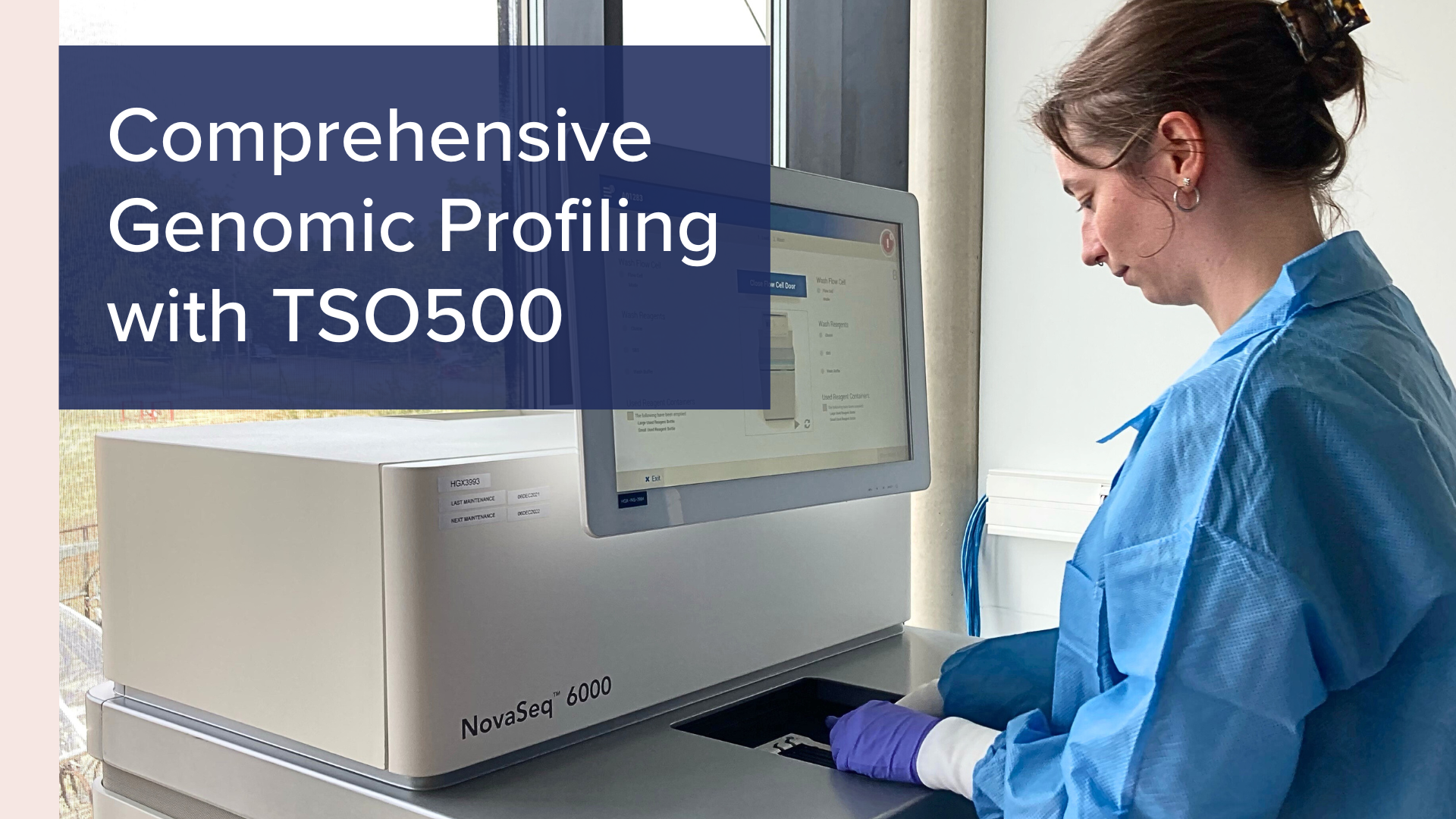
When conducting large-scale genomic studies, it is important to have access to required facilities and a reliable team with expertise in the latest technologies. Our global footprint and logistics expertise ensure that your samples reach our experts and undergo appropriate processing in a timely fashion. Large-scale genomic studies can become quite costly, which is why we offer the NovaSeq platform in conjunction with TSO500 to allow for analysis of 16-192 samples per run, thus providing scalable throughput for dynamic study sizes4.

Regulatory and Compliance Information

The TruSight Oncology Comprehensive panel (TSOComp) is approved by regulatory authorities both in Europe and United States, meaning it was validated under stringent regulatory standards for clinical diagnostics.

This regulatory approval ensures that TSOComp meets the necessary criteria for accuracy, reliability, and clinical utility in these locations.

Its compliance with global regulatory standards is crucial for its integration into clinical workflows, allowing the genomic data generated by TSOComp to inform critical treatment decisions with confidence.

At CellCarta, we offer our broad range of services and biomarker expertise to provide solutions for your study needs and to support our partners in propelling precision medicine. Contact our team to speak to an expert about your clinical trials.

About the author:

author photo

Sara Diels (PhD) is an assay development scientist at the Genomics Services unit within CellCarta. She led the implementation of the TSO500 High-Throughput and ctDNA assay at the Antwerp site. Her background is in molecular biology, and she has experience in the field of disease genetics where different -omics technologies are used to examine multifactorial inheritance. At CellCarta, Sara has expanded her expertise with the development and validation of PCR- and NGS-based assays.

References

  1. TruSight Oncology 500 Assay | For pan-cancer biomarkers in DNA and RNA. https://www.illumina.com/products/by-type/clinical-research-products/trusight-oncology-500.html.
  2. Pestinger, V. et al. Use of an Integrated Pan-Cancer Oncology Enrichment Next-Generation Sequencing Assay to Measure Tumour Mutational Burden and Detect Clinically Actionable Variants. Mol Diagn Ther 24, 339–349 (2020).
  3. Wei, B. et al. Evaluation of the TruSight Oncology 500 Assay for Routine Clinical Testing of Tumor Mutational Burden and Clinical Utility for Predicting Response to Pembrolizumab. Journal of Molecular Diagnostics 24, 600–608 (2022).
  4. NovaSeq 6000 System. https://www.illumina.com/systems/sequencing-platforms/novaseq.html

Genomic Profiling with TSO500: Advance Cancer Research Tool by CellCarta

May 15, 2023

Precise and reliable genomic profiling is key for identifying new therapeutic targets, making treatment decisions, and monitoring disease progression. Uncovering critical genetic information about tumor biology, predictive biomarkers, and immune responses can aid in the selection of appropriate patients and optimize study outcomes.

The TruSight Oncology 500 (TSO500) panel is a cutting-edge next-generation sequencing (NGS) tool that enables comprehensive genomic profiling across a broad range of cancer types.

It covers 523 cancer-related genes from DNA and 55 from RNA, allowing for the detection of single nucleotide variants (SNVs), insertions/deletions (indels), copy number variations (CNVs), and gene fusions.1

With to possibility to analyze both tissue and liquid biopsy samples, TSO500 can provide a holistic view of the tumor’s genetic landscape, aiding in the identification of therapeutic targets.

With TSO500, labs have the flexibility to choose between manual or automated workflows. However, automated workflows achieve high-quality results with higher throughput, enhancing lab efficiency and saving labor costs. TSO500 assays improve biomarker discovery and provide insightful details using only a minimal amount of sample, thus saving precious samples.

Additionally, TSO500 offers automation options, enabling labs to process samples with minimal hands-on time, making it a valuable tool in high-throughput environments. This automation feature allows for processing up to 192 samples per run, significantly reducing costs and turnaround time for large-scale studies.

How can the TSO500 panel improve the development of cancer therapies?

The development of immunotherapies requires rigorous assessment of immuno-oncology biomarkers, including TMB and MSI, as well as other genetic markers such as RNA fusions, single nucleotide variant (SNV), insertions/deletions (indels), and copy number variation, all of which can be determined via TSO500 2,3.

MSI status has traditionally been evaluated via PCR and immunohistochemistry techniques. While such methods provide qualitative insight into MSI-stable or MSI-high status, the TSO500 assay evaluates 130 specific homopolymer MSI marker sites, producing a quantitative score for MSI status1. Additionally, determining consistent TMB values at low mutation levels can pose challenges when using smaller panels.

TSO500 overcomes this obstacle by seamlessly integrating genomic content with advanced algorithms, resulting in precise TMB estimates highly concordant with whole-exome studies (WES).

Its dual capability to assess both DNA and RNA in a single assay makes it an incredibly valuable technology when looking into uncover actionable mutations.

Consolidating multiple biomarker assays into one comprehensive NGS assay requires less sample, provides a quicker turnaround time, and significantly increases the chances of finding positive biomarkers1.

Improve TSO500 testing with CellCarta’s support

When conducting large-scale genomic studies, it is important to have access to required facilities and a reliable team with expertise in the latest technologies. Our global footprint and logistics expertise ensure that your samples reach our experts and undergo appropriate processing in a timely fashion. Large-scale genomic studies can become quite costly, which is why we offer the NovaSeq platform in conjunction with TSO500 to allow for analysis of 16-192 samples per run, thus providing scalable throughput for dynamic study sizes4.

Regulatory and Compliance Information

The TruSight Oncology Comprehensive panel (TSOComp) is approved by regulatory authorities both in Europe and United States, meaning it was validated under stringent regulatory standards for clinical diagnostics.

This regulatory approval ensures that TSOComp meets the necessary criteria for accuracy, reliability, and clinical utility in these locations.

Its compliance with global regulatory standards is crucial for its integration into clinical workflows, allowing the genomic data generated by TSOComp to inform critical treatment decisions with confidence.

At CellCarta, we offer our broad range of services and biomarker expertise to provide solutions for your study needs and to support our partners in propelling precision medicine. Contact our team to speak to an expert about your clinical trials.

About the author:

author photo

Sara Diels (PhD) is an assay development scientist at the Genomics Services unit within CellCarta. She led the implementation of the TSO500 High-Throughput and ctDNA assay at the Antwerp site. Her background is in molecular biology, and she has experience in the field of disease genetics where different -omics technologies are used to examine multifactorial inheritance. At CellCarta, Sara has expanded her expertise with the development and validation of PCR- and NGS-based assays.

References

  1. TruSight Oncology 500 Assay | For pan-cancer biomarkers in DNA and RNA. https://www.illumina.com/products/by-type/clinical-research-products/trusight-oncology-500.html.
  2. Pestinger, V. et al. Use of an Integrated Pan-Cancer Oncology Enrichment Next-Generation Sequencing Assay to Measure Tumour Mutational Burden and Detect Clinically Actionable Variants. Mol Diagn Ther 24, 339–349 (2020).
  3. Wei, B. et al. Evaluation of the TruSight Oncology 500 Assay for Routine Clinical Testing of Tumor Mutational Burden and Clinical Utility for Predicting Response to Pembrolizumab. Journal of Molecular Diagnostics 24, 600–608 (2022).
  4. NovaSeq 6000 System. https://www.illumina.com/systems/sequencing-platforms/novaseq.html