George Demetri, MD and Charles Saywers, MD: Chats with Our Scientific Advisory Board

August 28, 2026

George Demetri, MD and Charles Saywers, MD, members of CellCarta's Scientific Advisory Board, join Christopher Ung, our Chief Scientific Business Officer, for a conversation on advancements in immunotherapies, treating solid tumors, and what's next for the field.

George Demetri, MD and Charles Saywers, MD: Chats with Our Scientific Advisory Board

August 28, 2026

George Demetri, MD and Charles Saywers, MD, members of CellCarta's Scientific Advisory Board, join Christopher Ung, our Chief Scientific Business Officer, for a conversation on advancements in immunotherapies, treating solid tumors, and what's next for the field.

Carl June, MD: Chats with Our Scientific Advisory Board

August 28, 2026

Carl June, MD, and member of CellCarta's Scientific Advisory Board, join Christopher Ung, our Chief Scientific Business Officer, for a conversation on advancements in immunotherapies, why ex vivo CAR-Ts aren't going to disappear, and what's next for the field.

Carl June, MD: Chats with Our Scientific Advisory Board

August 28, 2026

Carl June, MD, and member of CellCarta's Scientific Advisory Board, join Christopher Ung, our Chief Scientific Business Officer, for a conversation on advancements in immunotherapies, why ex vivo CAR-Ts aren't going to disappear, and what's next for the field.

Fit-for-purpose validation and inter-laboratory cross-validation of a TBNK flow cytometry assay with improved B cell monitoring capabilities

April 14, 2026

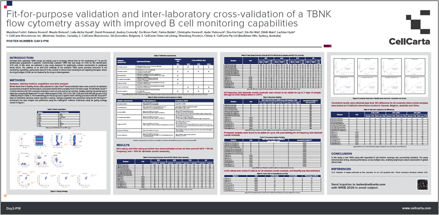
Fit-for-purpose validation and inter-laboratory cross-validation of a TBNK flow cytometry assay with improved B cell monitoring capabilities

Fit-for-purpose validation and inter-laboratory cross-validation of a TBNK flow cytometry assay with improved B cell monitoring capabilities

April 14, 2026

Fit-for-purpose validation and inter-laboratory cross-validation of a TBNK flow cytometry assay with improved B cell monitoring capabilities

TBNK Panels: How To Select the Optimal One

November 25, 2025

Flow cytometry–based TBNK panels are a mainstay of immune profiling, used to accurately enumerate T cells, B cells, and natural killer (NK) cells for a range of clinical purposes. Using the right TBNK panel configuration is essential for generating relevant results that support confident clinical decision-making, but identifying the most suitable option can be challenging.  

In this blog, we explore the key factors that influence TBNK panel selection and the importance of validated, standardized approaches in generating valuable immune profiling data

Why use a TBNK panel 

Accurate immune profiling is central to understanding the processes that influence how patients will respond to therapeutic interventions, and the reliability of those insights depends on consistent measurement of key lymphocyte populations. TBNK panels provide a standardized framework for generating that data, allowing immune responses to be compared across time points, patients, or clinical sites.  

By staining blood samples with antibodies that recognize cell surface markers, typically including CD3, CD4, CD8, CD19, and CD16/56, TBNK panels provide a high-level quantitative overview of a patient’s immune composition. Because different studies may focus on different immune subsets or endpoints, there are multiple TBNK panel configurations you can choose from. Each panel configuration provides distinct insights, and selecting the appropriate one ensures that the data generated is relevant and interpretable within the clinical context. 

How do you choose the right TBNK panel? 

Several factors influence which TBNK panel configuration is most appropriate for a given study or clinical application. 

Therapeutic target 

When and how a TBNK panel is deployed is largely determined by the type of therapy being investigated. Currently, TBNK analysis is most often used to track B-cell aplasia or T-cell expansion and contraction, applications that are particularly relevant to CAR-T and T-cell engager (TCE) therapies targeting B cells.  

Once the decision is made to use a TBNK assay, panel design is guided by the therapeutic target and the immune cell populations most relevant to the study. Standard off-the-shelf (OTS) configurations are often used as a foundation, but these can be adapted to specific study questions with additional markers.  

Regulatory and clinical context 

If data is to be included in regulatory submissions, an assay with the appropriate validation or IVD certification should be used to ensure compliance and data integrity. Other panel configurations may be validated for use in clinical settings, where they can support applications such as defining secondary endpoints or establishing patient onboarding and exclusion criteria.  

Turnaround time and workflow 

For clinical decision-making, rapid and standardized data delivery is also a key factor. An off-the-shelf, validated TBNK workflow allows for efficient processing and official reporting, enabling timely decisions such as the release of healthy donors based on B-cell repopulation following anti-B-cell therapy. 

Validated TBNK Panels from CellCarta 

Whatever your specific needs, CellCarta offers a portfolio of validated TBNK flow cytometry panels. Each panel has been validated in-house and cross-validated across CellCarta’s global sites to confirm reproducible performance between laboratories, with emphasis given to stability testing to ensure consistent performance over time. .  

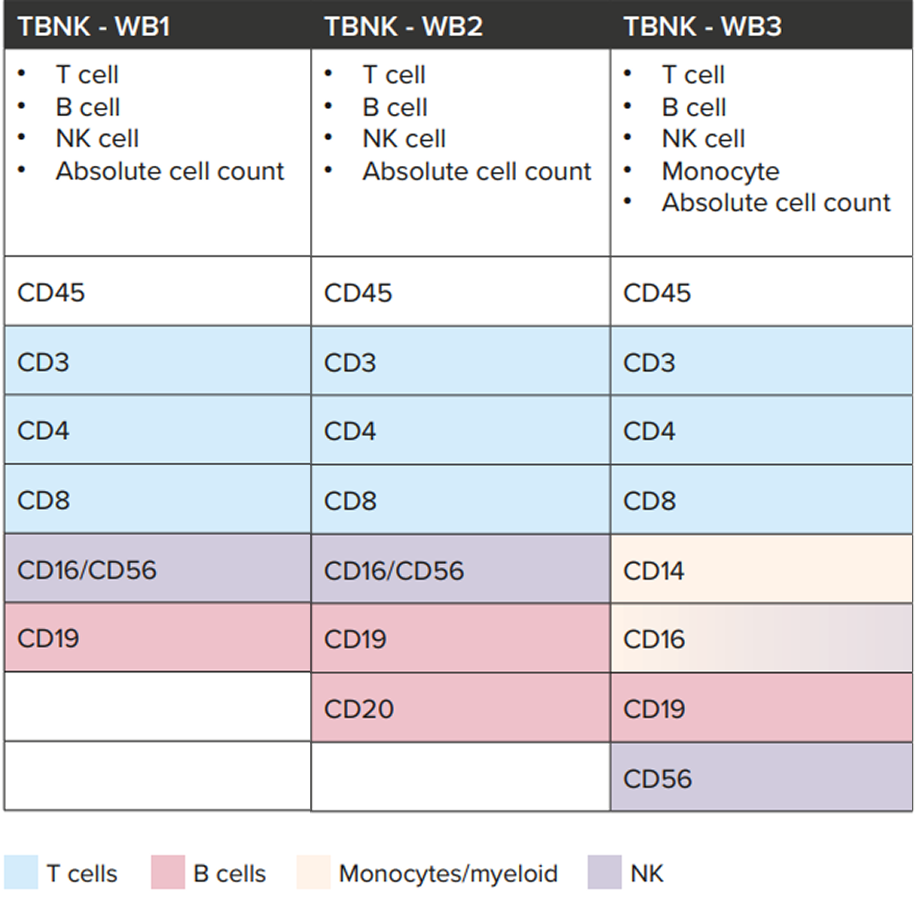
Several OTS TBNK configurations are available, each including a specific set of markers suited to different study objectives (Table 1): 

  • Standard TBNK panel (WB1): includes CD45, CD3, CD4, CD8, CD16/56, and CD19, providing broad coverage of T, B, and NK cells 
  • TBNK + CD20 panel (WB2): incorporates CD20 to better identify B-cell populations  
  • TBNK + monocytes panel (WB3): adds CD14, CD16, and CD56 to extend analysis to monocytes and NK-cell subsets  

These OTS configurations can be used as starting points that can be adapted to accommodate a vast range of specific research or clinical needs. Additional markers may be introduced to capture other immune populations, provided the panel size is kept small enough to remain compatible with the lyse/no-wash protocol.


Table 1Overview of CellCarta’s OTS TBNK panel configurations and marker composition. Each configuration can be further adapted with additional markers as required, within the limits of the lyse/no-wash protocol. 

Our standard TBNK panel (TBNK 1) has IVD status (when used according to the manufacturer’s specifications), making it suitable for use in regulatory submissions. Our other non-IVD panels are validated internally to the same rigorous standard and can be deployed to inform clinical decisions, such as patient onboarding or healthy donor release criteria. 

The absolute counts reported from the TBNK enumeration panels provide stand-alone values that enable direct comparison across studies and support consistent tracking of immune populations. Additionally, to support timely decision-making, CellCarta’s standardized TBNK workflows enable official reporting within five days, providing the speed required for responsive immune monitoring.  

Together, these measures ensure that CellCarta’s TBNK panels deliver high-quality, reproducible data that strengthens confidence in immune profiling results. 

Enable reliable results across TBNK assays 

Selecting the right TBNK panel depends on understanding how therapeutic target study goals, and clinical context shape immune monitoring requirements. Meeting those varied requirements calls for proven expertise in both assay development and implementation.  

Backed by deep experience in flow cytometry and assay validation, CellCarta can perform TBNK assays that combine reproducible performance, cross-site consistency, and efficient reporting. 

To find out more about how CellCarta’s TBNK panel expertise can support your research, talk to one of our experts.


About the author:

author photo

Céline Vandamme is a Scientific Business Director at CellCarta, specializing in the flow cytometry platform. With a PhD in immunology, and a broad expertise gained through her work at various academic and pharmaceutical institutions, Céline has profuse experience in designing flow cytometry assays to support immune monitoring activities in clinical trials.

TBNK Panels: How To Select the Optimal One

November 25, 2025

Flow cytometry–based TBNK panels are a mainstay of immune profiling, used to accurately enumerate T cells, B cells, and natural killer (NK) cells for a range of clinical purposes. Using the right TBNK panel configuration is essential for generating relevant results that support confident clinical decision-making, but identifying the most suitable option can be challenging.  

In this blog, we explore the key factors that influence TBNK panel selection and the importance of validated, standardized approaches in generating valuable immune profiling data

Why use a TBNK panel 

Accurate immune profiling is central to understanding the processes that influence how patients will respond to therapeutic interventions, and the reliability of those insights depends on consistent measurement of key lymphocyte populations. TBNK panels provide a standardized framework for generating that data, allowing immune responses to be compared across time points, patients, or clinical sites.  

By staining blood samples with antibodies that recognize cell surface markers, typically including CD3, CD4, CD8, CD19, and CD16/56, TBNK panels provide a high-level quantitative overview of a patient’s immune composition. Because different studies may focus on different immune subsets or endpoints, there are multiple TBNK panel configurations you can choose from. Each panel configuration provides distinct insights, and selecting the appropriate one ensures that the data generated is relevant and interpretable within the clinical context. 

How do you choose the right TBNK panel? 

Several factors influence which TBNK panel configuration is most appropriate for a given study or clinical application. 

Therapeutic target 

When and how a TBNK panel is deployed is largely determined by the type of therapy being investigated. Currently, TBNK analysis is most often used to track B-cell aplasia or T-cell expansion and contraction, applications that are particularly relevant to CAR-T and T-cell engager (TCE) therapies targeting B cells.  

Once the decision is made to use a TBNK assay, panel design is guided by the therapeutic target and the immune cell populations most relevant to the study. Standard off-the-shelf (OTS) configurations are often used as a foundation, but these can be adapted to specific study questions with additional markers.  

Regulatory and clinical context 

If data is to be included in regulatory submissions, an assay with the appropriate validation or IVD certification should be used to ensure compliance and data integrity. Other panel configurations may be validated for use in clinical settings, where they can support applications such as defining secondary endpoints or establishing patient onboarding and exclusion criteria.  

Turnaround time and workflow 

For clinical decision-making, rapid and standardized data delivery is also a key factor. An off-the-shelf, validated TBNK workflow allows for efficient processing and official reporting, enabling timely decisions such as the release of healthy donors based on B-cell repopulation following anti-B-cell therapy. 

Validated TBNK Panels from CellCarta 

Whatever your specific needs, CellCarta offers a portfolio of validated TBNK flow cytometry panels. Each panel has been validated in-house and cross-validated across CellCarta’s global sites to confirm reproducible performance between laboratories, with emphasis given to stability testing to ensure consistent performance over time. .  

Several OTS TBNK configurations are available, each including a specific set of markers suited to different study objectives (Table 1): 

  • Standard TBNK panel (WB1): includes CD45, CD3, CD4, CD8, CD16/56, and CD19, providing broad coverage of T, B, and NK cells 
  • TBNK + CD20 panel (WB2): incorporates CD20 to better identify B-cell populations  
  • TBNK + monocytes panel (WB3): adds CD14, CD16, and CD56 to extend analysis to monocytes and NK-cell subsets  

These OTS configurations can be used as starting points that can be adapted to accommodate a vast range of specific research or clinical needs. Additional markers may be introduced to capture other immune populations, provided the panel size is kept small enough to remain compatible with the lyse/no-wash protocol.


Table 1Overview of CellCarta’s OTS TBNK panel configurations and marker composition. Each configuration can be further adapted with additional markers as required, within the limits of the lyse/no-wash protocol. 

Our standard TBNK panel (TBNK 1) has IVD status (when used according to the manufacturer’s specifications), making it suitable for use in regulatory submissions. Our other non-IVD panels are validated internally to the same rigorous standard and can be deployed to inform clinical decisions, such as patient onboarding or healthy donor release criteria. 

The absolute counts reported from the TBNK enumeration panels provide stand-alone values that enable direct comparison across studies and support consistent tracking of immune populations. Additionally, to support timely decision-making, CellCarta’s standardized TBNK workflows enable official reporting within five days, providing the speed required for responsive immune monitoring.  

Together, these measures ensure that CellCarta’s TBNK panels deliver high-quality, reproducible data that strengthens confidence in immune profiling results. 

Enable reliable results across TBNK assays 

Selecting the right TBNK panel depends on understanding how therapeutic target study goals, and clinical context shape immune monitoring requirements. Meeting those varied requirements calls for proven expertise in both assay development and implementation.  

Backed by deep experience in flow cytometry and assay validation, CellCarta can perform TBNK assays that combine reproducible performance, cross-site consistency, and efficient reporting. 

To find out more about how CellCarta’s TBNK panel expertise can support your research, talk to one of our experts.


About the author:

author photo

Céline Vandamme is a Scientific Business Director at CellCarta, specializing in the flow cytometry platform. With a PhD in immunology, and a broad expertise gained through her work at various academic and pharmaceutical institutions, Céline has profuse experience in designing flow cytometry assays to support immune monitoring activities in clinical trials.

Tregs in Focus: How a Nobel-Winning Finding is Shaping Clinical Research

October 22, 2025

The 2025 Nobel Prize in Physiology or Medicine recognized discoveries that transformed our understanding of how the immune system maintains balance. Mary Brunkow, Frederick Ramsdell, and Shimon Sakaguchi were awarded for uncovering the mechanisms of peripheral immune tolerance, a process that prevents our immune system from unduly attacking the body’s own tissues.¹  

Through decades of research, they revealed that immune tolerance is not just established during T-cell development in the thymus but is actively maintained throughout life by a specialized population of FOXP3 regulatory T cells (Tregs). Their research demonstrated that Treg cells act as the immune system’s ‘braking mechanism’, suppressing inappropriate or excessive activation that could harm the body.¹  

Their discoveries bridged a gap that had long puzzled immunologists, explaining how the immune system is dynamically self-regulating throughout life to distinguish between harmful invaders and healthy cells. That knowledge paved the way to today’s wave of Treg-focused clinical trials, where researchers are investigating how tipping the immune balance one way or the other can be achieved through precise manipulation of Treg cells.   

What is the current focus of Treg clinical trials? 

Building on these foundational discoveries, current clinical research is focused on how modulating Treg activity can improve outcomes in patients with cancer and autoimmune diseases. 

Oncology 

In oncology, Treg clinical trials are exploring ways to reduce the suppressive activity of Tregs within the tumor microenvironment (TME), where these cells often accumulate and inhibit anti-tumor immunity.2

Key approaches under investigation include: 

  • Checkpoint and cytokine modulation, such as combining PD-(L)1 and VEGF inhibitors, which can alter the TME to reduce immune suppression, including Treg activity2
  • Blocking chemokine receptors, using antibodies that target CCR4 or CCR8 to prevent Tregs from migrating into the TME and deplete them2
  • Targeted depletion, including next-generation antibody–drug conjugates (ADCs) designed to selectively eliminate Tregs through markers such as CD253 

Early-phase studies show that these strategies can enhance cytotoxic T-cell activity and improve responses to checkpoint inhibitors.1,2  Further research is focused on improving selectivity, ensuring that Tregs in the TME can be modulated without disrupting the peripheral populations needed to maintain overall immune balance.2 

Autoimmune diseases 

In autoimmune and inflammatory disorders, clinical strategies take the opposite approach by enhancing or stabilizing Tregs to re-establish immune tolerance.  

Current research includes:  

  • Cytokine-based stimulation, using low-dose or engineered interleukin-2 (IL-2) therapies that selectively expand Tregs while avoiding activation of effector T cells4,5
  • Adoptive or engineered Treg therapies, such as CAR-Tregs and T-cell receptor (TCR)-Tregs, which are being developed to deliver site-specific immune suppression and promote graft tolerance in transplantation4,5 

Early clinical data suggest that these approaches can expand functional Tregs and reduce autoimmune inflammation without broadly suppressing protective immunity. As research progresses, the aim is to improve the scalability, safety, and efficacy of Treg cell therapies to make them more accessible to a broader patient population.5 

How are Tregs assessed in clinical trials?  

Accurate measurement of Tregs is essential for evaluating treatment effects in clinical studies. Assessments are typically performed in both tissue and blood samples, each providing complementary insight into Treg frequency, localization, and function. 

Assessment in tissues  

Tissue analysis provides spatial information about where Tregs are located and how they interact with other immune cells within the tissue microenvironment. Biomarkers such as FOXP3, CD3, and CD4 are commonly used to identify Tregs in histopathology samples. To capture this in high resolution, researchers can use multiplex imaging platforms such as the Lunaphore COMET™ system to detect dozens of markers at single-cell resolution, enabling high-plex visualization of Tregs and their surrounding context. 

Assessment in blood  

Blood-based analysis focuses on circulating Tregs, typically defined by CD25 and CD127 expression, with FOXP3 used as a confirmatory marker. Flow cytometry can be used to quantify Treg frequency and phenotype in whole blood or isolated peripheral blood mononuclear cells (PBMCs). 

What are the challenges of assessing Tregs in clinical trials?  

While these analytical methods are well established, accurately quantifying Tregs in clinical samples can be challenging. Their low frequency and sensitivity to handling, in addition to a reliance on intracellular markers such as FOXP3 mean that even minor differences in sample processing and methodology can impact results.  

Sample stability and processing 

Tregs are highly sensitive to sample handling, which can make them difficult to assess accurately . During PBMC isolation, there can be up to a fivefold decrease in Treg populations, even when samples are processed within 24 hours of blood draw.        

To avoid this, researchers can use CytoChex® blood collection tubes for whole blood collection if Tregs are a key readout, as this approach has been found to better preserve Treg cell frequency and marker expression over time.  

Staining resolution 

Because FOXP3 is an intracellular marker, accurate detection depends on optimized fixation, permeabilization, and gating. Inconsistent preparation may blur signal distinction and complicate gating. 

This can be addressed through optimized staining protocols and refined gating strategies, which can improve the reproducibility and clarity of FOXP3 Treg readouts in whole blood.  

Treg heterogeneity 

Tregs are not a uniform population. Differences in stability, activation state, and function can complicate data interpretation, particularly when bulk assays average out signals across diverse subsets. 

This complexity can be addressed through single-cell transcriptomic and TCR-sequencing approaches, which can identify distinct Treg subpopulations and track how individual clones shift in phenotype or functionality over time. These methods provide a more detailed view of Treg diversity and its relevance to therapeutic response. 

Looking ahead 

The discoveries recognized by the 2025 Nobel Prize underpin an expanding focus on Tregs in clinical immunology and oncology research. As therapeutic programs increasingly incorporate Treg assessment, advances in sample stabilization, staining workflows, and multi-omic profiling can help deliver more consistent, high-resolution data to help researchers characterize these cells and link their activity to clinical outcomes. 

These deeper insights can help pave the way for more precise, effective therapies that could offer new potential to treat cancer and autoimmune diseases. 

Contact us to find out how CellCarta’s immunology and biomarker expertise can support Treg assessment and analysis.  

About the author:

author photo

Céline Vandamme is a Scientific Business Director at CellCarta, specializing in the flow cytometry platform. With a PhD in immunology, and a broad expertise gained through her work at various academic and pharmaceutical institutions, Céline has profuse experience in designing flow cytometry assays to support immune monitoring activities in clinical trials.

References 

  1. https://www.nobelprize.org/prizes/medicine/2025/popular-information/ 
  2. https://pmc.ncbi.nlm.nih.gov/articles/PMC9588644/#:~:text=
    There%20are%20seven%20proposed%20methods,Treg%20cytokine%20secretion%2C%20(6)
      
  3. https://www.nature.com/articles/s12276-023-01080-3  
  4. https://www.frontiersin.org/journals/immunology/articles/10.3389/fimmu.2025.1511671/full  
  5. https://www.delveinsight.com/blog/treg-cell-based-therapies-in-pipeline 
Tregs in Focus: How a Nobel-Winning Finding is Shaping Clinical Research

October 22, 2025

The 2025 Nobel Prize in Physiology or Medicine recognized discoveries that transformed our understanding of how the immune system maintains balance. Mary Brunkow, Frederick Ramsdell, and Shimon Sakaguchi were awarded for uncovering the mechanisms of peripheral immune tolerance, a process that prevents our immune system from unduly attacking the body’s own tissues.¹  

Through decades of research, they revealed that immune tolerance is not just established during T-cell development in the thymus but is actively maintained throughout life by a specialized population of FOXP3 regulatory T cells (Tregs). Their research demonstrated that Treg cells act as the immune system’s ‘braking mechanism’, suppressing inappropriate or excessive activation that could harm the body.¹  

Their discoveries bridged a gap that had long puzzled immunologists, explaining how the immune system is dynamically self-regulating throughout life to distinguish between harmful invaders and healthy cells. That knowledge paved the way to today’s wave of Treg-focused clinical trials, where researchers are investigating how tipping the immune balance one way or the other can be achieved through precise manipulation of Treg cells.   

What is the current focus of Treg clinical trials? 

Building on these foundational discoveries, current clinical research is focused on how modulating Treg activity can improve outcomes in patients with cancer and autoimmune diseases. 

Oncology 

In oncology, Treg clinical trials are exploring ways to reduce the suppressive activity of Tregs within the tumor microenvironment (TME), where these cells often accumulate and inhibit anti-tumor immunity.2

Key approaches under investigation include: 

  • Checkpoint and cytokine modulation, such as combining PD-(L)1 and VEGF inhibitors, which can alter the TME to reduce immune suppression, including Treg activity2
  • Blocking chemokine receptors, using antibodies that target CCR4 or CCR8 to prevent Tregs from migrating into the TME and deplete them2
  • Targeted depletion, including next-generation antibody–drug conjugates (ADCs) designed to selectively eliminate Tregs through markers such as CD253 

Early-phase studies show that these strategies can enhance cytotoxic T-cell activity and improve responses to checkpoint inhibitors.1,2  Further research is focused on improving selectivity, ensuring that Tregs in the TME can be modulated without disrupting the peripheral populations needed to maintain overall immune balance.2 

Autoimmune diseases 

In autoimmune and inflammatory disorders, clinical strategies take the opposite approach by enhancing or stabilizing Tregs to re-establish immune tolerance.  

Current research includes:  

  • Cytokine-based stimulation, using low-dose or engineered interleukin-2 (IL-2) therapies that selectively expand Tregs while avoiding activation of effector T cells4,5
  • Adoptive or engineered Treg therapies, such as CAR-Tregs and T-cell receptor (TCR)-Tregs, which are being developed to deliver site-specific immune suppression and promote graft tolerance in transplantation4,5 

Early clinical data suggest that these approaches can expand functional Tregs and reduce autoimmune inflammation without broadly suppressing protective immunity. As research progresses, the aim is to improve the scalability, safety, and efficacy of Treg cell therapies to make them more accessible to a broader patient population.5 

How are Tregs assessed in clinical trials?  

Accurate measurement of Tregs is essential for evaluating treatment effects in clinical studies. Assessments are typically performed in both tissue and blood samples, each providing complementary insight into Treg frequency, localization, and function. 

Assessment in tissues  

Tissue analysis provides spatial information about where Tregs are located and how they interact with other immune cells within the tissue microenvironment. Biomarkers such as FOXP3, CD3, and CD4 are commonly used to identify Tregs in histopathology samples. To capture this in high resolution, researchers can use multiplex imaging platforms such as the Lunaphore COMET™ system to detect dozens of markers at single-cell resolution, enabling high-plex visualization of Tregs and their surrounding context. 

Assessment in blood  

Blood-based analysis focuses on circulating Tregs, typically defined by CD25 and CD127 expression, with FOXP3 used as a confirmatory marker. Flow cytometry can be used to quantify Treg frequency and phenotype in whole blood or isolated peripheral blood mononuclear cells (PBMCs). 

What are the challenges of assessing Tregs in clinical trials?  

While these analytical methods are well established, accurately quantifying Tregs in clinical samples can be challenging. Their low frequency and sensitivity to handling, in addition to a reliance on intracellular markers such as FOXP3 mean that even minor differences in sample processing and methodology can impact results.  

Sample stability and processing 

Tregs are highly sensitive to sample handling, which can make them difficult to assess accurately . During PBMC isolation, there can be up to a fivefold decrease in Treg populations, even when samples are processed within 24 hours of blood draw.        

To avoid this, researchers can use CytoChex® blood collection tubes for whole blood collection if Tregs are a key readout, as this approach has been found to better preserve Treg cell frequency and marker expression over time.  

Staining resolution 

Because FOXP3 is an intracellular marker, accurate detection depends on optimized fixation, permeabilization, and gating. Inconsistent preparation may blur signal distinction and complicate gating. 

This can be addressed through optimized staining protocols and refined gating strategies, which can improve the reproducibility and clarity of FOXP3 Treg readouts in whole blood.  

Treg heterogeneity 

Tregs are not a uniform population. Differences in stability, activation state, and function can complicate data interpretation, particularly when bulk assays average out signals across diverse subsets. 

This complexity can be addressed through single-cell transcriptomic and TCR-sequencing approaches, which can identify distinct Treg subpopulations and track how individual clones shift in phenotype or functionality over time. These methods provide a more detailed view of Treg diversity and its relevance to therapeutic response. 

Looking ahead 

The discoveries recognized by the 2025 Nobel Prize underpin an expanding focus on Tregs in clinical immunology and oncology research. As therapeutic programs increasingly incorporate Treg assessment, advances in sample stabilization, staining workflows, and multi-omic profiling can help deliver more consistent, high-resolution data to help researchers characterize these cells and link their activity to clinical outcomes. 

These deeper insights can help pave the way for more precise, effective therapies that could offer new potential to treat cancer and autoimmune diseases. 

Contact us to find out how CellCarta’s immunology and biomarker expertise can support Treg assessment and analysis.  

About the author:

author photo

Céline Vandamme is a Scientific Business Director at CellCarta, specializing in the flow cytometry platform. With a PhD in immunology, and a broad expertise gained through her work at various academic and pharmaceutical institutions, Céline has profuse experience in designing flow cytometry assays to support immune monitoring activities in clinical trials.

References 

  1. https://www.nobelprize.org/prizes/medicine/2025/popular-information/ 
  2. https://pmc.ncbi.nlm.nih.gov/articles/PMC9588644/#:~:text=
    There%20are%20seven%20proposed%20methods,Treg%20cytokine%20secretion%2C%20(6)
      
  3. https://www.nature.com/articles/s12276-023-01080-3  
  4. https://www.frontiersin.org/journals/immunology/articles/10.3389/fimmu.2025.1511671/full  
  5. https://www.delveinsight.com/blog/treg-cell-based-therapies-in-pipeline