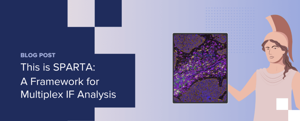
This is SPARTA: A Framework for Multiplex Immunofluorescence Analysis

November 13, 2025
What is spatial biology? 

Understanding how cells interact in their native context is critical for the development of novel therapeutics and effective diagnostics. Conventional assays or single-cell approaches often miss this layer of information, either by averaging signals across the tissue or by removing cells from their tissue environment. Spatial biology, on the other hand, preserves tissue architecture, allowing researchers to study cell type, location, and interaction together to reveal a more complete picture of disease biology.  

The value of spatial biology  

Spatial biology provides researchers with insights that can directly inform therapeutic development and translational decision-making. By combining cellular detail with spatial context, it enables teams to: 

  • Map the tissue microenvironment to understand how e.g. immune, stromal, and cancer cells shape disease progression 
  • Identify new therapeutic targets and validate them in situ, rather than in dissociated systems 
  • Pinpoint mechanisms of treatment response or resistance by showing how spatial relationships can influence therapeutic effects, giving insight into why patients with seemingly similar profiles can respond differently 
  • Strengthen biomarker discovery and validation by identifying spatial patterns that correlate with clinical outcomes 

Spatial biology methods generate exceptionally rich datasets that capture multiple layers of tissue complexity. One of the more common spatial biology approaches is multiplex immunofluorescence (mIF), which enables multiple markers to be visualized on a single tissue section. While these datasets bring enormous value, their size and complexity can make them difficult to interpret. When multiple imaging platforms are in use, workflows can become fragmented, and researchers risk generating data that cannot easily be integrated, slowing discovery and translation. 

Realizing the full value of spatial biology requires standardized multiplex immunofluorescence protocols and structured workflows that can handle complex datasets and deliver clear, interpretable insights.  

SPARTA: Bringing order to spatial biology  

To simplify mIF workflows, CellCarta developed SPARTA (Spatial Phenotyping and Analysis in Regions of Tissue with AI)—a platform-agnostic framework that standardizes how datasets are processed and analyzed across imaging systems.  

SPARTA incorporates:  

  • Whole-slide imaging and QC across multiple platforms (Lunaphore Comet, Akoya PhenoImager, and Zeiss Axioscan Z1), offering flexibility and shorter lead times  
  • Segmentation and classification, using expert-designed, AI-enabled algorithms for segmentation, followed by single-cell feature extraction and rule-based classification 
  • Data export and advanced analytics, including summary outputs (densities, proportions), object-based single-cell data with coordinates, and advanced analyses such as supervised/unsupervised phenotyping, proximity analysis, and cell neighborhood mapping 
  • Custom data delivery that avoids generic “data dumps,” providing instead tailored insights aligned with specific research questions  

By combining platform flexibility, advanced analytics, and tailored reporting, the SPARTA framework ensures that data from different platforms is processed consistently, so researchers can more easily extract meaningful patterns from complex datasets.

Drive discovery with spatial biology  

By revealing the organization of complex tissue environments and capturing cellular interactions, spatial biology continues to provide critical insights that shape drug development. mIF makes it possible to generate rich, multi-dimensional datasets that inform preclinical research and translational decisions. With structured workflows like SPARTA, supported by strong expertise and collaborations across assay development, pathology, imaging, and data science teams, this data can be transformed into tailored outputs that can help accelerate discovery, strengthen biomarker programs, and support clinical development. 

To find out more about how our SPARTA framework could support your spatial biology workflows, get in touch with one of our experts! 


About the author

author photo

Yannick Waumans is Executive Director of Histopathology Operations at CellCarta, where he oversees the Histopathology Lab Services, Technical Transfer Office, and Digital Pathology Solutions. With a PhD in Pharmaceutical Sciences and over a decade of experience in histopathology, imaging, and image analysis, Yannick is an expert in advancing and operationalizing digital pathology technologies.  

He is especially passionate about leveraging digital pathology to accelerate research and improve clinical outcomes. 
This is SPARTA: A Framework for Multiplex Immunofluorescence Analysis

November 13, 2025

What is spatial biology? 

Understanding how cells interact in their native context is critical for the development of novel therapeutics and effective diagnostics. Conventional assays or single-cell approaches often miss this layer of information, either by averaging signals across the tissue or by removing cells from their tissue environment. Spatial biology, on the other hand, preserves tissue architecture, allowing researchers to study cell type, location, and interaction together to reveal a more complete picture of disease biology.  

The value of spatial biology  

Spatial biology provides researchers with insights that can directly inform therapeutic development and translational decision-making. By combining cellular detail with spatial context, it enables teams to: 

  • Map the tissue microenvironment to understand how e.g. immune, stromal, and cancer cells shape disease progression 
  • Identify new therapeutic targets and validate them in situ, rather than in dissociated systems 
  • Pinpoint mechanisms of treatment response or resistance by showing how spatial relationships can influence therapeutic effects, giving insight into why patients with seemingly similar profiles can respond differently 
  • Strengthen biomarker discovery and validation by identifying spatial patterns that correlate with clinical outcomes 

Spatial biology methods generate exceptionally rich datasets that capture multiple layers of tissue complexity. One of the more common spatial biology approaches is multiplex immunofluorescence (mIF), which enables multiple markers to be visualized on a single tissue section. While these datasets bring enormous value, their size and complexity can make them difficult to interpret. When multiple imaging platforms are in use, workflows can become fragmented, and researchers risk generating data that cannot easily be integrated, slowing discovery and translation. 

Realizing the full value of spatial biology requires standardized multiplex immunofluorescence protocols and structured workflows that can handle complex datasets and deliver clear, interpretable insights.  

SPARTA: Bringing order to spatial biology  

To simplify mIF workflows, CellCarta developed SPARTA (Spatial Phenotyping and Analysis in Regions of Tissue with AI)—a platform-agnostic framework that standardizes how datasets are processed and analyzed across imaging systems.  

SPARTA incorporates:  

  • Whole-slide imaging and QC across multiple platforms (Lunaphore Comet, Akoya PhenoImager, and Zeiss Axioscan Z1), offering flexibility and shorter lead times  
  • Segmentation and classification, using expert-designed, AI-enabled algorithms for segmentation, followed by single-cell feature extraction and rule-based classification 
  • Data export and advanced analytics, including summary outputs (densities, proportions), object-based single-cell data with coordinates, and advanced analyses such as supervised/unsupervised phenotyping, proximity analysis, and cell neighborhood mapping 
  • Custom data delivery that avoids generic “data dumps,” providing instead tailored insights aligned with specific research questions  

By combining platform flexibility, advanced analytics, and tailored reporting, the SPARTA framework ensures that data from different platforms is processed consistently, so researchers can more easily extract meaningful patterns from complex datasets.

Drive discovery with spatial biology  

By revealing the organization of complex tissue environments and capturing cellular interactions, spatial biology continues to provide critical insights that shape drug development. mIF makes it possible to generate rich, multi-dimensional datasets that inform preclinical research and translational decisions. With structured workflows like SPARTA, supported by strong expertise and collaborations across assay development, pathology, imaging, and data science teams, this data can be transformed into tailored outputs that can help accelerate discovery, strengthen biomarker programs, and support clinical development. 

To find out more about how our SPARTA framework could support your spatial biology workflows, get in touch with one of our experts! 


About the author

author photo

Yannick Waumans is Executive Director of Histopathology Operations at CellCarta, where he oversees the Histopathology Lab Services, Technical Transfer Office, and Digital Pathology Solutions. With a PhD in Pharmaceutical Sciences and over a decade of experience in histopathology, imaging, and image analysis, Yannick is an expert in advancing and operationalizing digital pathology technologies.  

He is especially passionate about leveraging digital pathology to accelerate research and improve clinical outcomes. 

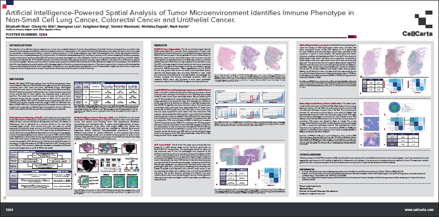
Artificial Intelligence-powered Spatial Analysis of Tumor Microenvironment Identifies Immune Phenotype in Non- Small Cell Lung Cancer, Colorectal Cancer and Urothelial Cancer.

October 30, 2025

Artificial Intelligence-Powered Spatial Analysis of Tumor Microenvironment Identifies Immune Phenotype in Non-Small Cell Lung Cancer, Colorectal Cancer and Urothelial Cancer.

Artificial Intelligence-powered Spatial Analysis of Tumor Microenvironment Identifies Immune Phenotype in Non- Small Cell Lung Cancer, Colorectal Cancer and Urothelial Cancer.

October 30, 2025

Artificial Intelligence-Powered Spatial Analysis of Tumor Microenvironment Identifies Immune Phenotype in Non-Small Cell Lung Cancer, Colorectal Cancer and Urothelial Cancer.

Poster Video: Induction of an effective immune response is an essential element of cancer immunotherapy

September 2, 2025

One way to identify the patients likely to respond to immunotherapy is by identifying the distribution of inflammatory cells relative to tumor cells. However, in spite of the compelling association of immune phenotypes with clinical outcome, there are not yet standard definitions or scoring methods.

See how our scientists developed a method for pathologists to identify desert, excluded and inflamed immune phenotypes in our new video!

Want more details? Read the poster!

Poster Video: Induction of an effective immune response is an essential element of cancer immunotherapy

September 2, 2025

One way to identify the patients likely to respond to immunotherapy is by identifying the distribution of inflammatory cells relative to tumor cells. However, in spite of the compelling association of immune phenotypes with clinical outcome, there are not yet standard definitions or scoring methods.

See how our scientists developed a method for pathologists to identify desert, excluded and inflamed immune phenotypes in our new video!

Want more details? Read the poster!

Biomarkers & AI in Future ADCs: Dr. Powles’ Insights

March 17, 2025

Antibody-drug conjugates (ADCs) are changing cancer treatment for the better, combining precision-targeting of cancer cells with the potency of chemotherapy.  

Perhaps one of the most exciting recent advances in ADCs was seen in a trial led by renowned oncologist Dr Thomas Powles. The combination treatment of enfortumab vedotin (EV), a nectin-4 ADC, and pembrolizumab (pembro), a PD-1 inhibitor, more than doubled the median survival of metastatic bladder cancer patients, from one year with standard chemotherapy, to two and a half years.   

Thanks to these transformative results, EV-pembro has superseded traditional platinum-based chemotherapy as the first-line treatment, and Dr Powles believes a cure for bladder cancer is now possible—something that would have seemed unimaginable just a few years ago.  

In our latest Let’s Talk webcast, we had the privilege of talking to Dr Powles about the future possibilities of ADCs. While in our previous blog we discussed his thoughts on the future of ADCs in cancer treatment, here we summarize his insights on the pivotal role that biomarkers and AI could play in their development.  

The importance of biomarkers in future ADC development

As with many other new precision medicine therapeutic strategies, a key hurdle in ADC development is understanding why some patients benefit more than others. While EV-pembro is broadly effective in treating bladder cancer, since nectin-4 is expressed in 90% of the cancer cells, there are still some patients that show limited benefit, and understanding why has proven challenging. Identifying biomarkers to stratify subtle differences in patient profiles may be crucial to achieving better outcomes.  

“If we’re going to cure bladder cancer, we may not do so with EV-pembro alone,” said Dr Powles. “While it could serve as a baseline treatment for many, we must understand why some patients don’t respond as well, and how we can improve their response, if we are going to achieve a cure.”  

“That’s where the second generation of biomarkers come in“ he continues. “Using transcriptomics and multiplex analysis, we could see what’s expressed in non-responders and find out whether we should be using other agents or more complex immune therapy for those patients.” 

Dr Powles believes that biomarkers will be key to ADC breakthroughs beyond bladder cancer, too. “Deep down, I think the transformative result we’ve seen with EV-pembro in bladder cancer is not a black swan event,” he said. “I think it’s possible in subsets of other cancers.” 

While other ADCs have shown promise, such as those directed at TROP2 and HER2, patient selection criteria remain imprecise, confounding their real effectiveness.  

For TROP2 ADCs, such as sacituzumab govitecan, despite rapid progress, a more refined biomarker strategy is still needed to identify the patients who will benefit the most from this treatment. Similarly, with the HER2-low breast cancer ADC trastuzumab deruxtecan, there is debate around how HER2 expression levels correlate with response rates. HER2 scoring methods can produce inconsistent results, therefore more precise biomarker assessment could substantially improve treatment outcomes.

The role of AI in future ADC development

To help in advancing ADC development, researchers are looking to leverage AI technologies to improve biomarker assessment. Traditional pathology methods rely on human interpretation, which can lead to inconsistencies in how biomarkers are assessed across different labs and clinical settings.

“Diagnostic pathology using AI can bring a standardization that currently isn’t possible with traditional methods,” said Powles. “So far, we haven’t been overly successful with biomarker development. To move forwards, we need AI technology to reduce variability and improve accuracy in patient selection.”

Currently, researchers are investigating the use of AI in identifying responders and non-responders for TROP2 ADCs. When it comes to their use in lung cancer treatment, Dr Powles states: “If you can find the 30% of patients that have a strong response, that could potentially be transformative. If we’re not currently seeing any improvement over traditional chemotherapy with TROP2 ADCs, then we need to find a smarter way of assessing biomarkers, and that could be through AI.”

Looking ahead

The future of ADCs holds great promise, with refined biomarker assessment and AI potentially playing a pivotal role in the development of more personalized therapies in finely-stratified patient cohorts.  

In the full webcast, Dr Powles shares his thoughts on the current and future treatment landscape of ADCs, including where he believes there is most potential for another breakthrough ADC treatment, and the technologies and strategies that could drive their development.  

Don’t miss out on hearing the firsthand insights of a true ADC expert—watch the webcast on demand today: Webcast – Thomas Powles – Gated | CellCarta.

About the author:

author photo

Céline Vandamme is a Scientific Business Director at CellCarta, specializing in the flow cytometry platform. With a PhD in immunology, and a broad expertise gained through her work at various academic and pharmaceutical institutions, Céline has profuse experience in designing flow cytometry assays to support immune monitoring activities in clinical trials.

Biomarkers & AI in Future ADCs: Dr. Powles’ Insights

March 17, 2025

Antibody-drug conjugates (ADCs) are changing cancer treatment for the better, combining precision-targeting of cancer cells with the potency of chemotherapy.  

Perhaps one of the most exciting recent advances in ADCs was seen in a trial led by renowned oncologist Dr Thomas Powles. The combination treatment of enfortumab vedotin (EV), a nectin-4 ADC, and pembrolizumab (pembro), a PD-1 inhibitor, more than doubled the median survival of metastatic bladder cancer patients, from one year with standard chemotherapy, to two and a half years.   

Thanks to these transformative results, EV-pembro has superseded traditional platinum-based chemotherapy as the first-line treatment, and Dr Powles believes a cure for bladder cancer is now possible—something that would have seemed unimaginable just a few years ago.  

In our latest Let’s Talk webcast, we had the privilege of talking to Dr Powles about the future possibilities of ADCs. While in our previous blog we discussed his thoughts on the future of ADCs in cancer treatment, here we summarize his insights on the pivotal role that biomarkers and AI could play in their development.  

The importance of biomarkers in future ADC development

As with many other new precision medicine therapeutic strategies, a key hurdle in ADC development is understanding why some patients benefit more than others. While EV-pembro is broadly effective in treating bladder cancer, since nectin-4 is expressed in 90% of the cancer cells, there are still some patients that show limited benefit, and understanding why has proven challenging. Identifying biomarkers to stratify subtle differences in patient profiles may be crucial to achieving better outcomes.  

“If we’re going to cure bladder cancer, we may not do so with EV-pembro alone,” said Dr Powles. “While it could serve as a baseline treatment for many, we must understand why some patients don’t respond as well, and how we can improve their response, if we are going to achieve a cure.”  

“That’s where the second generation of biomarkers come in“ he continues. “Using transcriptomics and multiplex analysis, we could see what’s expressed in non-responders and find out whether we should be using other agents or more complex immune therapy for those patients.” 

Dr Powles believes that biomarkers will be key to ADC breakthroughs beyond bladder cancer, too. “Deep down, I think the transformative result we’ve seen with EV-pembro in bladder cancer is not a black swan event,” he said. “I think it’s possible in subsets of other cancers.” 

While other ADCs have shown promise, such as those directed at TROP2 and HER2, patient selection criteria remain imprecise, confounding their real effectiveness.  

For TROP2 ADCs, such as sacituzumab govitecan, despite rapid progress, a more refined biomarker strategy is still needed to identify the patients who will benefit the most from this treatment. Similarly, with the HER2-low breast cancer ADC trastuzumab deruxtecan, there is debate around how HER2 expression levels correlate with response rates. HER2 scoring methods can produce inconsistent results, therefore more precise biomarker assessment could substantially improve treatment outcomes.

The role of AI in future ADC development

To help in advancing ADC development, researchers are looking to leverage AI technologies to improve biomarker assessment. Traditional pathology methods rely on human interpretation, which can lead to inconsistencies in how biomarkers are assessed across different labs and clinical settings.

“Diagnostic pathology using AI can bring a standardization that currently isn’t possible with traditional methods,” said Powles. “So far, we haven’t been overly successful with biomarker development. To move forwards, we need AI technology to reduce variability and improve accuracy in patient selection.”

Currently, researchers are investigating the use of AI in identifying responders and non-responders for TROP2 ADCs. When it comes to their use in lung cancer treatment, Dr Powles states: “If you can find the 30% of patients that have a strong response, that could potentially be transformative. If we’re not currently seeing any improvement over traditional chemotherapy with TROP2 ADCs, then we need to find a smarter way of assessing biomarkers, and that could be through AI.”

Looking ahead

The future of ADCs holds great promise, with refined biomarker assessment and AI potentially playing a pivotal role in the development of more personalized therapies in finely-stratified patient cohorts.  

In the full webcast, Dr Powles shares his thoughts on the current and future treatment landscape of ADCs, including where he believes there is most potential for another breakthrough ADC treatment, and the technologies and strategies that could drive their development.  

Don’t miss out on hearing the firsthand insights of a true ADC expert—watch the webcast on demand today: Webcast – Thomas Powles – Gated | CellCarta.

About the author:

author photo

Céline Vandamme is a Scientific Business Director at CellCarta, specializing in the flow cytometry platform. With a PhD in immunology, and a broad expertise gained through her work at various academic and pharmaceutical institutions, Céline has profuse experience in designing flow cytometry assays to support immune monitoring activities in clinical trials.

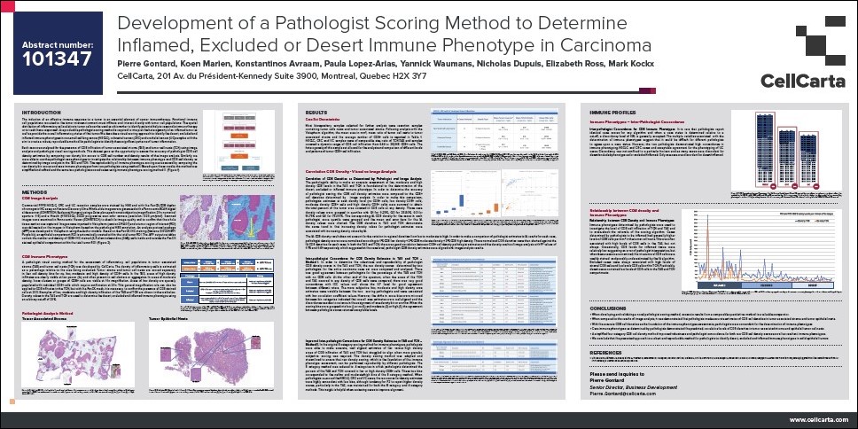
Development of a Pathologist Scoring Method to Determine Inflamed, Excluded or Desert Immune Phenotype in Carcinoma

March 5, 2025

Development of a Pathologist Scoring Method to Determine Inflamed, Excluded or Desert Immune Phenotype in Carcinoma

scoring metiodd to determine inflamed, excluded or desert Immune Phenotype in Carcinoma

Development of a Pathologist Scoring Method to Determine Inflamed, Excluded or Desert Immune Phenotype in Carcinoma

March 5, 2025

Development of a Pathologist Scoring Method to Determine Inflamed, Excluded or Desert Immune Phenotype in Carcinoma

scoring metiodd to determine inflamed, excluded or desert Immune Phenotype in Carcinoma