April 10, 2026
CellCarta has deep expertise in developing flow cytometry panels for clinical use and provides the highest standard of quality with CAP accreditated and CLIA certified sites.
To support the monitoring of all immune cell populations, we catered a list of specific panels for both your whole blood and cryopreserved PBMC samples.
Off-the-shelf (OTS) panels were pre-developed and validated at all global sites with the same level of validation and provide the fastest runway to clinical sample analysis, and catalogue panels were pre-developed and validated at specific sites and can be rapidly deployed upon request.
April 10, 2026
CellCarta has deep expertise in developing flow cytometry panels for clinical use and provides the highest standard of quality with CAP accreditated and CLIA certified sites.
To support the monitoring of all immune cell populations, we catered a list of specific panels for both your whole blood and cryopreserved PBMC samples.
Off-the-shelf (OTS) panels were pre-developed and validated at all global sites with the same level of validation and provide the fastest runway to clinical sample analysis, and catalogue panels were pre-developed and validated at specific sites and can be rapidly deployed upon request.
November 25, 2025
Flow cytometry–based TBNK panels are a mainstay of immune profiling, used to accurately enumerate T cells, B cells, and natural killer (NK) cells for a range of clinical purposes. Using the right TBNK panel configuration is essential for generating relevant results that support confident clinical decision-making, but identifying the most suitable option can be challenging.
In this blog, we explore the key factors that influence TBNK panel selection and the importance of validated, standardized approaches in generating valuable immune profiling data
Accurate immune profiling is central to understanding the processes that influence how patients will respond to therapeutic interventions, and the reliability of those insights depends on consistent measurement of key lymphocyte populations. TBNK panels provide a standardized framework for generating that data, allowing immune responses to be compared across time points, patients, or clinical sites.
By staining blood samples with antibodies that recognize cell surface markers, typically including CD3, CD4, CD8, CD19, and CD16/56, TBNK panels provide a high-level quantitative overview of a patient’s immune composition. Because different studies may focus on different immune subsets or endpoints, there are multiple TBNK panel configurations you can choose from. Each panel configuration provides distinct insights, and selecting the appropriate one ensures that the data generated is relevant and interpretable within the clinical context.
Several factors influence which TBNK panel configuration is most appropriate for a given study or clinical application.
When and how a TBNK panel is deployed is largely determined by the type of therapy being investigated. Currently, TBNK analysis is most often used to track B-cell aplasia or T-cell expansion and contraction, applications that are particularly relevant to CAR-T and T-cell engager (TCE) therapies targeting B cells.
Once the decision is made to use a TBNK assay, panel design is guided by the therapeutic target and the immune cell populations most relevant to the study. Standard off-the-shelf (OTS) configurations are often used as a foundation, but these can be adapted to specific study questions with additional markers.
If data is to be included in regulatory submissions, an assay with the appropriate validation or IVD certification should be used to ensure compliance and data integrity. Other panel configurations may be validated for use in clinical settings, where they can support applications such as defining secondary endpoints or establishing patient onboarding and exclusion criteria.
For clinical decision-making, rapid and standardized data delivery is also a key factor. An off-the-shelf, validated TBNK workflow allows for efficient processing and official reporting, enabling timely decisions such as the release of healthy donors based on B-cell repopulation following anti-B-cell therapy.
Validated TBNK Panels from CellCarta
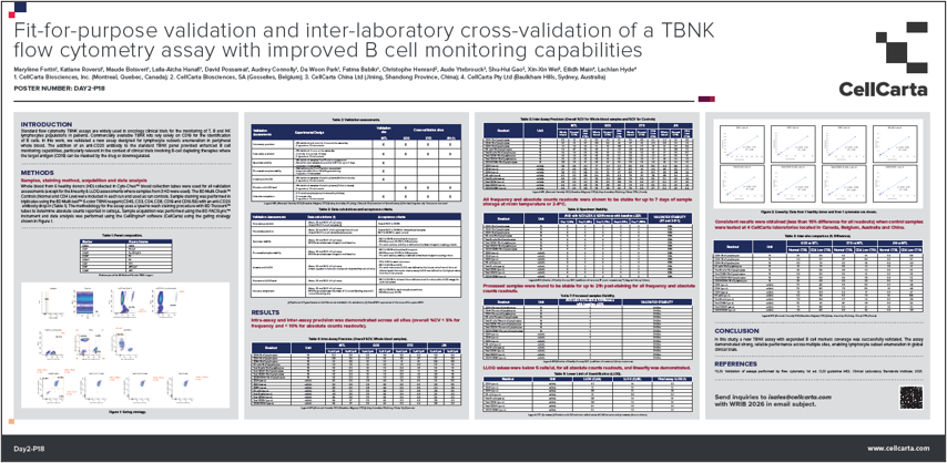
Whatever your specific needs, CellCarta offers a portfolio of validated TBNK flow cytometry panels. Each panel has been validated in-house and cross-validated across CellCarta’s global sites to confirm reproducible performance between laboratories, with emphasis given to stability testing to ensure consistent performance over time. .
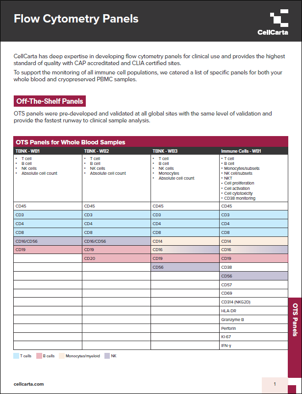
Several OTS TBNK configurations are available, each including a specific set of markers suited to different study objectives (Table 1):
These OTS configurations can be used as starting points that can be adapted to accommodate a vast range of specific research or clinical needs. Additional markers may be introduced to capture other immune populations, provided the panel size is kept small enough to remain compatible with the lyse/no-wash protocol.
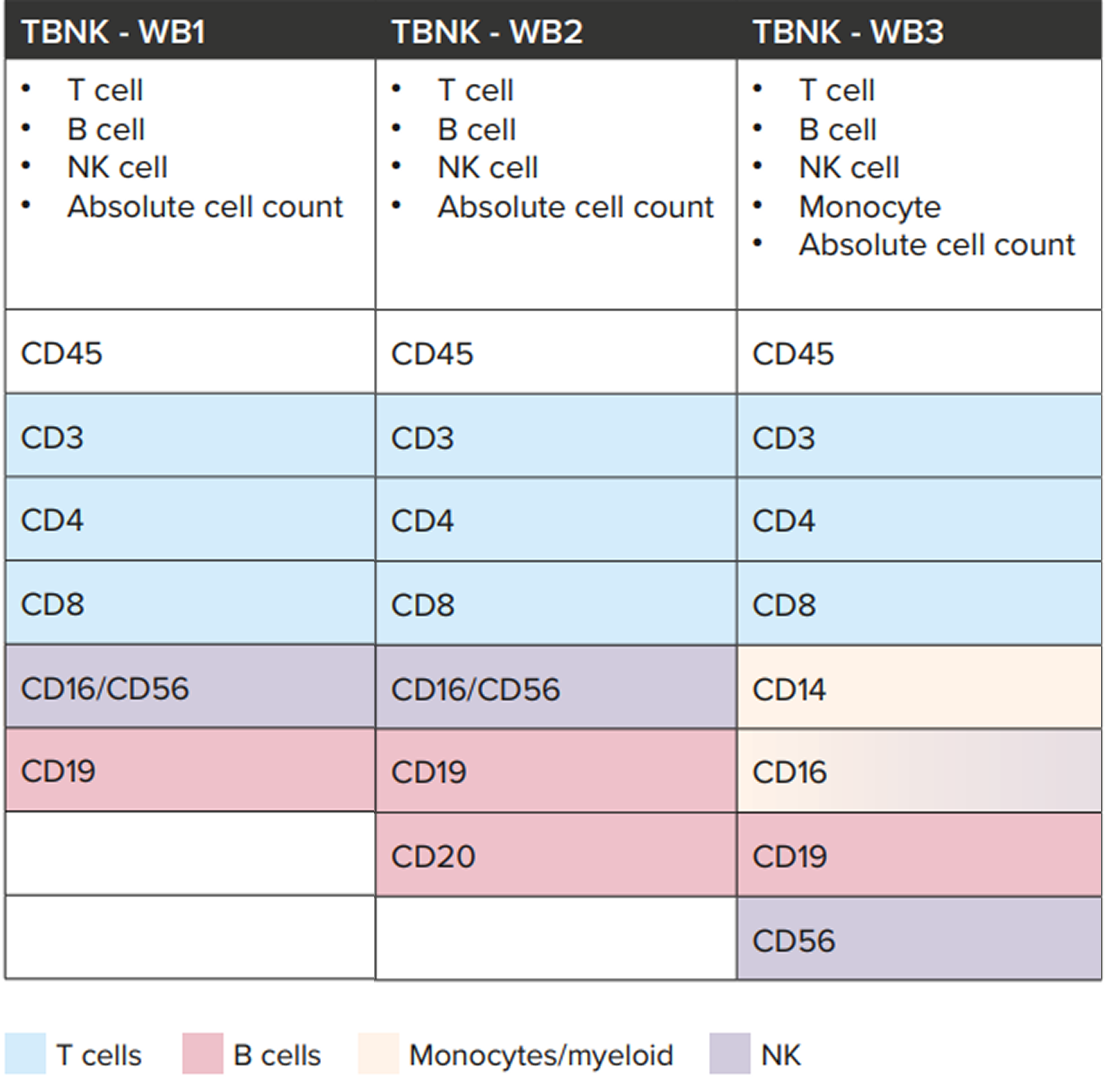
Table 1: Overview of CellCarta’s OTS TBNK panel configurations and marker composition. Each configuration can be further adapted with additional markers as required, within the limits of the lyse/no-wash protocol.
Our standard TBNK panel (TBNK 1) has IVD status (when used according to the manufacturer’s specifications), making it suitable for use in regulatory submissions. Our other non-IVD panels are validated internally to the same rigorous standard and can be deployed to inform clinical decisions, such as patient onboarding or healthy donor release criteria.
The absolute counts reported from the TBNK enumeration panels provide stand-alone values that enable direct comparison across studies and support consistent tracking of immune populations. Additionally, to support timely decision-making, CellCarta’s standardized TBNK workflows enable official reporting within five days, providing the speed required for responsive immune monitoring.
Together, these measures ensure that CellCarta’s TBNK panels deliver high-quality, reproducible data that strengthens confidence in immune profiling results.
Selecting the right TBNK panel depends on understanding how therapeutic target study goals, and clinical context shape immune monitoring requirements. Meeting those varied requirements calls for proven expertise in both assay development and implementation.
Backed by deep experience in flow cytometry and assay validation, CellCarta can perform TBNK assays that combine reproducible performance, cross-site consistency, and efficient reporting.
To find out more about how CellCarta’s TBNK panel expertise can support your research, talk to one of our experts.
About the author:

Céline Vandamme is a Scientific Business Director at CellCarta, specializing in the flow cytometry platform. With a PhD in immunology, and a broad expertise gained through her work at various academic and pharmaceutical institutions, Céline has profuse experience in designing flow cytometry assays to support immune monitoring activities in clinical trials.
November 25, 2025
Flow cytometry–based TBNK panels are a mainstay of immune profiling, used to accurately enumerate T cells, B cells, and natural killer (NK) cells for a range of clinical purposes. Using the right TBNK panel configuration is essential for generating relevant results that support confident clinical decision-making, but identifying the most suitable option can be challenging.
In this blog, we explore the key factors that influence TBNK panel selection and the importance of validated, standardized approaches in generating valuable immune profiling data
Accurate immune profiling is central to understanding the processes that influence how patients will respond to therapeutic interventions, and the reliability of those insights depends on consistent measurement of key lymphocyte populations. TBNK panels provide a standardized framework for generating that data, allowing immune responses to be compared across time points, patients, or clinical sites.
By staining blood samples with antibodies that recognize cell surface markers, typically including CD3, CD4, CD8, CD19, and CD16/56, TBNK panels provide a high-level quantitative overview of a patient’s immune composition. Because different studies may focus on different immune subsets or endpoints, there are multiple TBNK panel configurations you can choose from. Each panel configuration provides distinct insights, and selecting the appropriate one ensures that the data generated is relevant and interpretable within the clinical context.
Several factors influence which TBNK panel configuration is most appropriate for a given study or clinical application.
When and how a TBNK panel is deployed is largely determined by the type of therapy being investigated. Currently, TBNK analysis is most often used to track B-cell aplasia or T-cell expansion and contraction, applications that are particularly relevant to CAR-T and T-cell engager (TCE) therapies targeting B cells.
Once the decision is made to use a TBNK assay, panel design is guided by the therapeutic target and the immune cell populations most relevant to the study. Standard off-the-shelf (OTS) configurations are often used as a foundation, but these can be adapted to specific study questions with additional markers.
If data is to be included in regulatory submissions, an assay with the appropriate validation or IVD certification should be used to ensure compliance and data integrity. Other panel configurations may be validated for use in clinical settings, where they can support applications such as defining secondary endpoints or establishing patient onboarding and exclusion criteria.
For clinical decision-making, rapid and standardized data delivery is also a key factor. An off-the-shelf, validated TBNK workflow allows for efficient processing and official reporting, enabling timely decisions such as the release of healthy donors based on B-cell repopulation following anti-B-cell therapy.
Validated TBNK Panels from CellCarta
Whatever your specific needs, CellCarta offers a portfolio of validated TBNK flow cytometry panels. Each panel has been validated in-house and cross-validated across CellCarta’s global sites to confirm reproducible performance between laboratories, with emphasis given to stability testing to ensure consistent performance over time. .
Several OTS TBNK configurations are available, each including a specific set of markers suited to different study objectives (Table 1):
These OTS configurations can be used as starting points that can be adapted to accommodate a vast range of specific research or clinical needs. Additional markers may be introduced to capture other immune populations, provided the panel size is kept small enough to remain compatible with the lyse/no-wash protocol.
Table 1: Overview of CellCarta’s OTS TBNK panel configurations and marker composition. Each configuration can be further adapted with additional markers as required, within the limits of the lyse/no-wash protocol.
Our standard TBNK panel (TBNK 1) has IVD status (when used according to the manufacturer’s specifications), making it suitable for use in regulatory submissions. Our other non-IVD panels are validated internally to the same rigorous standard and can be deployed to inform clinical decisions, such as patient onboarding or healthy donor release criteria.
The absolute counts reported from the TBNK enumeration panels provide stand-alone values that enable direct comparison across studies and support consistent tracking of immune populations. Additionally, to support timely decision-making, CellCarta’s standardized TBNK workflows enable official reporting within five days, providing the speed required for responsive immune monitoring.
Together, these measures ensure that CellCarta’s TBNK panels deliver high-quality, reproducible data that strengthens confidence in immune profiling results.
Selecting the right TBNK panel depends on understanding how therapeutic target study goals, and clinical context shape immune monitoring requirements. Meeting those varied requirements calls for proven expertise in both assay development and implementation.
Backed by deep experience in flow cytometry and assay validation, CellCarta can perform TBNK assays that combine reproducible performance, cross-site consistency, and efficient reporting.
To find out more about how CellCarta’s TBNK panel expertise can support your research, talk to one of our experts.
About the author:
Céline Vandamme is a Scientific Business Director at CellCarta, specializing in the flow cytometry platform. With a PhD in immunology, and a broad expertise gained through her work at various academic and pharmaceutical institutions, Céline has profuse experience in designing flow cytometry assays to support immune monitoring activities in clinical trials.
October 22, 2025
The 2025 Nobel Prize in Physiology or Medicine recognized discoveries that transformed our understanding of how the immune system maintains balance. Mary Brunkow, Frederick Ramsdell, and Shimon Sakaguchi were awarded for uncovering the mechanisms of peripheral immune tolerance, a process that prevents our immune system from unduly attacking the body’s own tissues.¹
Through decades of research, they revealed that immune tolerance is not just established during T-cell development in the thymus but is actively maintained throughout life by a specialized population of FOXP3⁺ regulatory T cells (Tregs). Their research demonstrated that Treg cells act as the immune system’s ‘braking mechanism’, suppressing inappropriate or excessive activation that could harm the body.¹
Their discoveries bridged a gap that had long puzzled immunologists, explaining how the immune system is dynamically self-regulating throughout life to distinguish between harmful invaders and healthy cells. That knowledge paved the way to today’s wave of Treg-focused clinical trials, where researchers are investigating how tipping the immune balance one way or the other can be achieved through precise manipulation of Treg cells.
Building on these foundational discoveries, current clinical research is focused on how modulating Treg activity can improve outcomes in patients with cancer and autoimmune diseases.
In oncology, Treg clinical trials are exploring ways to reduce the suppressive activity of Tregs within the tumor microenvironment (TME), where these cells often accumulate and inhibit anti-tumor immunity.2
Key approaches under investigation include:
Early-phase studies show that these strategies can enhance cytotoxic T-cell activity and improve responses to checkpoint inhibitors.1,2 Further research is focused on improving selectivity, ensuring that Tregs in the TME can be modulated without disrupting the peripheral populations needed to maintain overall immune balance.2
In autoimmune and inflammatory disorders, clinical strategies take the opposite approach by enhancing or stabilizing Tregs to re-establish immune tolerance.
Current research includes:
Early clinical data suggest that these approaches can expand functional Tregs and reduce autoimmune inflammation without broadly suppressing protective immunity. As research progresses, the aim is to improve the scalability, safety, and efficacy of Treg cell therapies to make them more accessible to a broader patient population.5
Accurate measurement of Tregs is essential for evaluating treatment effects in clinical studies. Assessments are typically performed in both tissue and blood samples, each providing complementary insight into Treg frequency, localization, and function.
Tissue analysis provides spatial information about where Tregs are located and how they interact with other immune cells within the tissue microenvironment. Biomarkers such as FOXP3, CD3, and CD4 are commonly used to identify Tregs in histopathology samples. To capture this in high resolution, researchers can use multiplex imaging platforms such as the Lunaphore COMET™ system to detect dozens of markers at single-cell resolution, enabling high-plex visualization of Tregs and their surrounding context.
Blood-based analysis focuses on circulating Tregs, typically defined by CD25 and CD127 expression, with FOXP3 used as a confirmatory marker. Flow cytometry can be used to quantify Treg frequency and phenotype in whole blood or isolated peripheral blood mononuclear cells (PBMCs).
While these analytical methods are well established, accurately quantifying Tregs in clinical samples can be challenging. Their low frequency and sensitivity to handling, in addition to a reliance on intracellular markers such as FOXP3 mean that even minor differences in sample processing and methodology can impact results.
Tregs are highly sensitive to sample handling, which can make them difficult to assess accurately . During PBMC isolation, there can be up to a fivefold decrease in Treg populations, even when samples are processed within 24 hours of blood draw.
To avoid this, researchers can use CytoChex® blood collection tubes for whole blood collection if Tregs are a key readout, as this approach has been found to better preserve Treg cell frequency and marker expression over time.
Because FOXP3 is an intracellular marker, accurate detection depends on optimized fixation, permeabilization, and gating. Inconsistent preparation may blur signal distinction and complicate gating.
This can be addressed through optimized staining protocols and refined gating strategies, which can improve the reproducibility and clarity of FOXP3⁺ Treg readouts in whole blood.
Tregs are not a uniform population. Differences in stability, activation state, and function can complicate data interpretation, particularly when bulk assays average out signals across diverse subsets.
This complexity can be addressed through single-cell transcriptomic and TCR-sequencing approaches, which can identify distinct Treg subpopulations and track how individual clones shift in phenotype or functionality over time. These methods provide a more detailed view of Treg diversity and its relevance to therapeutic response.
The discoveries recognized by the 2025 Nobel Prize underpin an expanding focus on Tregs in clinical immunology and oncology research. As therapeutic programs increasingly incorporate Treg assessment, advances in sample stabilization, staining workflows, and multi-omic profiling can help deliver more consistent, high-resolution data to help researchers characterize these cells and link their activity to clinical outcomes.
These deeper insights can help pave the way for more precise, effective therapies that could offer new potential to treat cancer and autoimmune diseases.
Contact us to find out how CellCarta’s immunology and biomarker expertise can support Treg assessment and analysis.
About the author:

Céline Vandamme is a Scientific Business Director at CellCarta, specializing in the flow cytometry platform. With a PhD in immunology, and a broad expertise gained through her work at various academic and pharmaceutical institutions, Céline has profuse experience in designing flow cytometry assays to support immune monitoring activities in clinical trials.
October 22, 2025
The 2025 Nobel Prize in Physiology or Medicine recognized discoveries that transformed our understanding of how the immune system maintains balance. Mary Brunkow, Frederick Ramsdell, and Shimon Sakaguchi were awarded for uncovering the mechanisms of peripheral immune tolerance, a process that prevents our immune system from unduly attacking the body’s own tissues.¹
Through decades of research, they revealed that immune tolerance is not just established during T-cell development in the thymus but is actively maintained throughout life by a specialized population of FOXP3⁺ regulatory T cells (Tregs). Their research demonstrated that Treg cells act as the immune system’s ‘braking mechanism’, suppressing inappropriate or excessive activation that could harm the body.¹
Their discoveries bridged a gap that had long puzzled immunologists, explaining how the immune system is dynamically self-regulating throughout life to distinguish between harmful invaders and healthy cells. That knowledge paved the way to today’s wave of Treg-focused clinical trials, where researchers are investigating how tipping the immune balance one way or the other can be achieved through precise manipulation of Treg cells.
Building on these foundational discoveries, current clinical research is focused on how modulating Treg activity can improve outcomes in patients with cancer and autoimmune diseases.
In oncology, Treg clinical trials are exploring ways to reduce the suppressive activity of Tregs within the tumor microenvironment (TME), where these cells often accumulate and inhibit anti-tumor immunity.2
Key approaches under investigation include:
Early-phase studies show that these strategies can enhance cytotoxic T-cell activity and improve responses to checkpoint inhibitors.1,2 Further research is focused on improving selectivity, ensuring that Tregs in the TME can be modulated without disrupting the peripheral populations needed to maintain overall immune balance.2
In autoimmune and inflammatory disorders, clinical strategies take the opposite approach by enhancing or stabilizing Tregs to re-establish immune tolerance.
Current research includes:
Early clinical data suggest that these approaches can expand functional Tregs and reduce autoimmune inflammation without broadly suppressing protective immunity. As research progresses, the aim is to improve the scalability, safety, and efficacy of Treg cell therapies to make them more accessible to a broader patient population.5
Accurate measurement of Tregs is essential for evaluating treatment effects in clinical studies. Assessments are typically performed in both tissue and blood samples, each providing complementary insight into Treg frequency, localization, and function.
Tissue analysis provides spatial information about where Tregs are located and how they interact with other immune cells within the tissue microenvironment. Biomarkers such as FOXP3, CD3, and CD4 are commonly used to identify Tregs in histopathology samples. To capture this in high resolution, researchers can use multiplex imaging platforms such as the Lunaphore COMET™ system to detect dozens of markers at single-cell resolution, enabling high-plex visualization of Tregs and their surrounding context.
Blood-based analysis focuses on circulating Tregs, typically defined by CD25 and CD127 expression, with FOXP3 used as a confirmatory marker. Flow cytometry can be used to quantify Treg frequency and phenotype in whole blood or isolated peripheral blood mononuclear cells (PBMCs).
While these analytical methods are well established, accurately quantifying Tregs in clinical samples can be challenging. Their low frequency and sensitivity to handling, in addition to a reliance on intracellular markers such as FOXP3 mean that even minor differences in sample processing and methodology can impact results.
Tregs are highly sensitive to sample handling, which can make them difficult to assess accurately . During PBMC isolation, there can be up to a fivefold decrease in Treg populations, even when samples are processed within 24 hours of blood draw.
To avoid this, researchers can use CytoChex® blood collection tubes for whole blood collection if Tregs are a key readout, as this approach has been found to better preserve Treg cell frequency and marker expression over time.
Because FOXP3 is an intracellular marker, accurate detection depends on optimized fixation, permeabilization, and gating. Inconsistent preparation may blur signal distinction and complicate gating.
This can be addressed through optimized staining protocols and refined gating strategies, which can improve the reproducibility and clarity of FOXP3⁺ Treg readouts in whole blood.
Tregs are not a uniform population. Differences in stability, activation state, and function can complicate data interpretation, particularly when bulk assays average out signals across diverse subsets.
This complexity can be addressed through single-cell transcriptomic and TCR-sequencing approaches, which can identify distinct Treg subpopulations and track how individual clones shift in phenotype or functionality over time. These methods provide a more detailed view of Treg diversity and its relevance to therapeutic response.
The discoveries recognized by the 2025 Nobel Prize underpin an expanding focus on Tregs in clinical immunology and oncology research. As therapeutic programs increasingly incorporate Treg assessment, advances in sample stabilization, staining workflows, and multi-omic profiling can help deliver more consistent, high-resolution data to help researchers characterize these cells and link their activity to clinical outcomes.
These deeper insights can help pave the way for more precise, effective therapies that could offer new potential to treat cancer and autoimmune diseases.
Contact us to find out how CellCarta’s immunology and biomarker expertise can support Treg assessment and analysis.
About the author:
Céline Vandamme is a Scientific Business Director at CellCarta, specializing in the flow cytometry platform. With a PhD in immunology, and a broad expertise gained through her work at various academic and pharmaceutical institutions, Céline has profuse experience in designing flow cytometry assays to support immune monitoring activities in clinical trials.
September 19, 2025
B cells are central to both protective immunity and autoimmune pathology. With the recent shift of cell therapies from oncology indications to autoimmune diseases, interest has been growing towards this immune cell population.1,2
Flow cytometry remains the gold standard for monitoring B cells in both research and clinical settings. However, lack of convention for classification as well as reliable and stable markers, especially following thawing of samples, can make it difficult to capture rare B-cell populations relevant to autoimmune drug development.
For translational research teams, this creates an urgent need for tools that deliver the sensitivity and consistency required to fully understand B-cell dynamics and drive the development of new B-cell therapies.
B cells are far from uniform. Transitional, naïve, memory, double-negative, and plasma cell subsets each play distinct roles in health and disease, but their phenotypic profiles often overlap. Inconsistent marker usage and variability in gating strategies have made it difficult to classify B-cell subsets consistently, which can complicate the comparison of results across studies.3
For drug developers, the lack of standardization creates real-world hurdles. Autoimmune therapies that aim to induce a B-cell reset (eliminating autoreactive populations and allowing the immune system to repopulate with naïve B cells) depend on precise monitoring of which subsets return after the B-cell aplasia phase. Without consistent, high-resolution phenotyping, it is difficult to determine whether a therapy is resetting B-cell populations in a way that supports durable disease control.
Recent efforts to harmonize approaches are beginning to address these issues, with publications recommending standardized marker sets to improve reproducibility and resolution of B-cell subsets.³ Markers such as CD21, once regarded mainly for their functional role, are now recognized as important phenotypic indicators that help distinguish distinct populations. Incorporating markers like these into flow cytometry panels is essential to provide the clarity and consistency needed for research on B-cell reset in autoimmune diseases.
To address these challenges, CellCarta has advanced its flow cytometry capabilities with a refined B-cell assay that improves the sensitivity and consistency of B-cell subset profiling.
We refined our panel strategy by incorporating markers such as CD21 as phenotypic indicators alongside other established markers. This enables further stratification of B-cell subtypes, providing better insight into both activated and developmentally distinct populations that are increasingly linked to autoimmune disease.
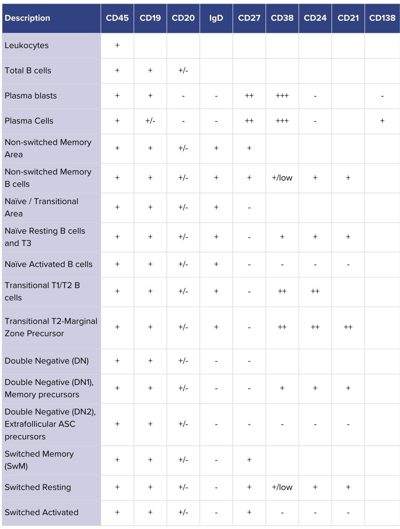
Table 1: B-cell subsets identifiable with CellCarta’s refined flow cytometry panel. Rows show major populations, while columns list the markers used to classify them. CD21 provides added resolution by distinguishing activated and developmentally distinct subsets relevant to autoimmune disease.
We’ve developed a refined bulk lysis method using the B-cell panel. In comparison to the direct whole blood (WB) analysis (100ul of blood), the improved bulk lysis method resulted in a 10-fold increase in the cell input compared to the traditional analysis using WB for flow cytometry assay (Figure 1). As more cells are acquired, rare populations can now be assessed with better precision. The whole method was evaluated at both 4°C and room temperature (RT), providing flexibility for the sample management and transportation.
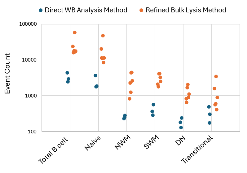
Figure 1: Event counts of total B cells and subsets acquired from healthy donors using direct whole blood analysis method (blue) and refined bulk lysis method (orange) WB – Whole Blood; NWM – Non-switched memory; SWM – Switched memory, DN – Double negative
To offer global site-to-site consistency, we switched our B-cell assay from the Fortessa to the Lyric platform, leveraging the built-in capabilities of this platform to achieve high inter-instrument/inter-site standardization. Unified workflows and directly comparable data make it easier to scale studies internationally, reduce variability, and build confidence in results across multicenter trials.
With refinements to its gating strategy, bulk lysis method, and global platform standardization, CellCarta’s B-cell flow cytometry assay provides the resolution, sensitivity, and reproducibility needed to study B-cell dynamics in detail. In the context of the development of B-cell reset therapies, these advancements enable the precise monitoring and consistency needed to better define which B-cell populations are depleted, which repopulate, and how these shifts influence autoimmune disease outcomes.
In addition to our B-cell panel, CellCarta can support researchers with both off-the-shelf panels for rapid deployment and custom panel development tailored to specific scientific questions. With this flexibility, pharma teams can choose the right approach for their needs, supporting clearer decision-making in the development of new B–cell–targeted therapies.

Alex Guo, PhD, is a Principal Scientist at CellCarta, specializing in immune monitoring and proteomics. With a doctorate in Immunology and Molecular Oncology, he has extensive expertise in developing and validating novel assays to address complex clinical needs. Alex has led immune monitoring analyses across multiple clinical programs, translating high-dimensional data into actionable insights for clients. He combines deep scientific expertise with practical project execution to advance translational and clinical research.
September 19, 2025
B cells are central to both protective immunity and autoimmune pathology. With the recent shift of cell therapies from oncology indications to autoimmune diseases, interest has been growing towards this immune cell population.1,2
Flow cytometry remains the gold standard for monitoring B cells in both research and clinical settings. However, lack of convention for classification as well as reliable and stable markers, especially following thawing of samples, can make it difficult to capture rare B-cell populations relevant to autoimmune drug development.
For translational research teams, this creates an urgent need for tools that deliver the sensitivity and consistency required to fully understand B-cell dynamics and drive the development of new B-cell therapies.
B cells are far from uniform. Transitional, naïve, memory, double-negative, and plasma cell subsets each play distinct roles in health and disease, but their phenotypic profiles often overlap. Inconsistent marker usage and variability in gating strategies have made it difficult to classify B-cell subsets consistently, which can complicate the comparison of results across studies.3
For drug developers, the lack of standardization creates real-world hurdles. Autoimmune therapies that aim to induce a B-cell reset (eliminating autoreactive populations and allowing the immune system to repopulate with naïve B cells) depend on precise monitoring of which subsets return after the B-cell aplasia phase. Without consistent, high-resolution phenotyping, it is difficult to determine whether a therapy is resetting B-cell populations in a way that supports durable disease control.
Recent efforts to harmonize approaches are beginning to address these issues, with publications recommending standardized marker sets to improve reproducibility and resolution of B-cell subsets.³ Markers such as CD21, once regarded mainly for their functional role, are now recognized as important phenotypic indicators that help distinguish distinct populations. Incorporating markers like these into flow cytometry panels is essential to provide the clarity and consistency needed for research on B-cell reset in autoimmune diseases.
To address these challenges, CellCarta has advanced its flow cytometry capabilities with a refined B-cell assay that improves the sensitivity and consistency of B-cell subset profiling.
We refined our panel strategy by incorporating markers such as CD21 as phenotypic indicators alongside other established markers. This enables further stratification of B-cell subtypes, providing better insight into both activated and developmentally distinct populations that are increasingly linked to autoimmune disease.
Table 1: B-cell subsets identifiable with CellCarta’s refined flow cytometry panel. Rows show major populations, while columns list the markers used to classify them. CD21 provides added resolution by distinguishing activated and developmentally distinct subsets relevant to autoimmune disease.
We’ve developed a refined bulk lysis method using the B-cell panel. In comparison to the direct whole blood (WB) analysis (100ul of blood), the improved bulk lysis method resulted in a 10-fold increase in the cell input compared to the traditional analysis using WB for flow cytometry assay (Figure 1). As more cells are acquired, rare populations can now be assessed with better precision. The whole method was evaluated at both 4°C and room temperature (RT), providing flexibility for the sample management and transportation.
Figure 1: Event counts of total B cells and subsets acquired from healthy donors using direct whole blood analysis method (blue) and refined bulk lysis method (orange) WB – Whole Blood; NWM – Non-switched memory; SWM – Switched memory, DN – Double negative
To offer global site-to-site consistency, we switched our B-cell assay from the Fortessa to the Lyric platform, leveraging the built-in capabilities of this platform to achieve high inter-instrument/inter-site standardization. Unified workflows and directly comparable data make it easier to scale studies internationally, reduce variability, and build confidence in results across multicenter trials.
With refinements to its gating strategy, bulk lysis method, and global platform standardization, CellCarta’s B-cell flow cytometry assay provides the resolution, sensitivity, and reproducibility needed to study B-cell dynamics in detail. In the context of the development of B-cell reset therapies, these advancements enable the precise monitoring and consistency needed to better define which B-cell populations are depleted, which repopulate, and how these shifts influence autoimmune disease outcomes.
In addition to our B-cell panel, CellCarta can support researchers with both off-the-shelf panels for rapid deployment and custom panel development tailored to specific scientific questions. With this flexibility, pharma teams can choose the right approach for their needs, supporting clearer decision-making in the development of new B–cell–targeted therapies.
Alex Guo, PhD, is a Principal Scientist at CellCarta, specializing in immune monitoring and proteomics. With a doctorate in Immunology and Molecular Oncology, he has extensive expertise in developing and validating novel assays to address complex clinical needs. Alex has led immune monitoring analyses across multiple clinical programs, translating high-dimensional data into actionable insights for clients. He combines deep scientific expertise with practical project execution to advance translational and clinical research.