Optimizing Cell Therapy with Advanced VCN and Cell Counting

July 16, 2024
Optimizing Cell Therapy Clinical Testing

Cell therapy targets, constructs, and agents are expanding and diversifying — fast. Clinical testing must now comprehensively characterize an ever-broader number and array of therapies, while continuing to prioritize urgency and efficiency in their development and approval.

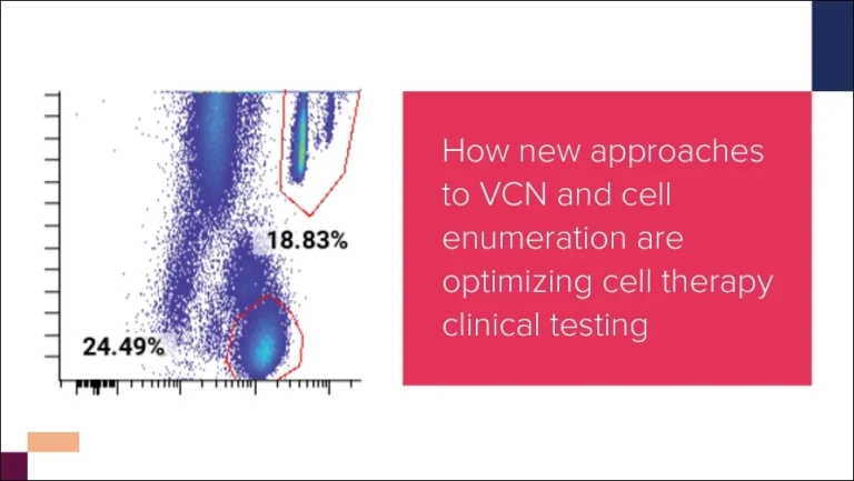
We delve into how clinical testing is evolving to meet demand in this rapidly changing landscape in our Cell Therapy Trends Report, and examine a few key areas where cell therapy testing programs are changing. One of these is cell enumeration and vector copy number (VCN) determination, which provide insights on how a given therapeutic construct is behaving in the body.

Below, we explore how techniques for cell enumeration and VCN determination are being adapted to assess greater numbers of increasingly complex and diversified therapy candidates, and discuss what the future holds for this key area of clinical testing.

Understanding cell enumeration and VCN in clinical testing

Cell enumeration and VCN determination methods assess cell number (via flow cytometric detection of surface proteins) and the number of constructs transduced into a cell (via PCR-based quantification of DNA), respectively. Together, the two measure cell therapy expansion and determine the abundance and persistence of therapeutic cells — crucial to ascertain how various cell types are responding to a therapy.

As cell therapies and sample types diversify, clinical testing for cell enumeration and VCN determination must assess a broader array of targets, requiring more specialized means of analysis. However, developers must also keep speed in mind, and balance specialization with efficiency to ensure that therapies are evaluated in a timely way.

So, what does the future hold for this realm of clinical testing?

Complementary methods

Cell enumeration and VCN determination techniques take complementary measurements from different angles, using different methods and analytes to evaluate cell therapies.

By utilizing both approaches in early-phase trials, cell therapy developers can gain high confidence in how they are interpreting their data, thereby producing a more reliable and robust picture of therapy expansion, and also lending confidence to each individual assay.

Alignment can also streamline testing. Cell enumeration via flow cytometry, despite providing additional phenotyping information on expanded cells, can be difficult to scale up, and so is often limited in later-stage use due to the challenges of collecting and transporting samples off-site. But, if aligned with cell enumeration in early-phase testing, VCN can act as a proxy measure of cell number during later-stage testing, simplifying logistics.

Increasing customization

As more diverse cell therapies reach clinical stages, cell enumeration and VCN determination methods need increasing customization — to track proprietary constructs as patients become more likely to receive multiple therapies, for instance. Additionally, new therapeutic cells such as CAR-macrophages may require novel antigen targets to be monitored in various tissues other than circulating blood, requiring new protocols for collection and analysis.

Enhancing modularity

Although customized assays are valuable, they are time- and resource-consuming to develop and validate. Modularization can minimize the timeline impact of assay customization. Testing programs for cell enumeration and VCN determination can be made quicker and easier to deploy by identifying generic analytes; and by utilizing modular data analytics to streamline readouts, and accelerate data processing.

Cell enumeration and VCN in the ever-changing cell therapy landscape

With the accelerating pace and increasing diversity of cell therapy development, clinical testing must become more responsive and adaptive, leveraging different approaches to deliver readouts that provide meaningful insights into cell therapy action and host response.

We dig deeper into emerging testing approaches for cell enumeration and VCN determination, and explore the future of cell therapy clinical testing more generally, in our Cell Therapy Trends Report. Download the report now, or contact our team to speak to an expert about your cell therapy needs.

About the author:

author photo

Laïla-Aïcha Hanafi is the Director of Scientific Laboratory Operations in flow cytometry at CellCarta. She supplemented her PhD in immuno-oncology with post-doctoral studies in translational biomarkers for cell therapies at the Fred Hutchinson Cancer Center. Laïla has combined scientific knowledge and operational efficiency to address biomarker needs in clinical trial and prioritizing high quality data to move therapies to the next stage of clinical deployment.


Optimizing Cell Therapy with Advanced VCN and Cell Counting

July 16, 2024

Optimizing Cell Therapy Clinical Testing

Cell therapy targets, constructs, and agents are expanding and diversifying — fast. Clinical testing must now comprehensively characterize an ever-broader number and array of therapies, while continuing to prioritize urgency and efficiency in their development and approval.

We delve into how clinical testing is evolving to meet demand in this rapidly changing landscape in our Cell Therapy Trends Report, and examine a few key areas where cell therapy testing programs are changing. One of these is cell enumeration and vector copy number (VCN) determination, which provide insights on how a given therapeutic construct is behaving in the body.

Below, we explore how techniques for cell enumeration and VCN determination are being adapted to assess greater numbers of increasingly complex and diversified therapy candidates, and discuss what the future holds for this key area of clinical testing.

Understanding cell enumeration and VCN in clinical testing

Cell enumeration and VCN determination methods assess cell number (via flow cytometric detection of surface proteins) and the number of constructs transduced into a cell (via PCR-based quantification of DNA), respectively. Together, the two measure cell therapy expansion and determine the abundance and persistence of therapeutic cells — crucial to ascertain how various cell types are responding to a therapy.

As cell therapies and sample types diversify, clinical testing for cell enumeration and VCN determination must assess a broader array of targets, requiring more specialized means of analysis. However, developers must also keep speed in mind, and balance specialization with efficiency to ensure that therapies are evaluated in a timely way.

So, what does the future hold for this realm of clinical testing?

Complementary methods

Cell enumeration and VCN determination techniques take complementary measurements from different angles, using different methods and analytes to evaluate cell therapies.

By utilizing both approaches in early-phase trials, cell therapy developers can gain high confidence in how they are interpreting their data, thereby producing a more reliable and robust picture of therapy expansion, and also lending confidence to each individual assay.

Alignment can also streamline testing. Cell enumeration via flow cytometry, despite providing additional phenotyping information on expanded cells, can be difficult to scale up, and so is often limited in later-stage use due to the challenges of collecting and transporting samples off-site. But, if aligned with cell enumeration in early-phase testing, VCN can act as a proxy measure of cell number during later-stage testing, simplifying logistics.

Increasing customization

As more diverse cell therapies reach clinical stages, cell enumeration and VCN determination methods need increasing customization — to track proprietary constructs as patients become more likely to receive multiple therapies, for instance. Additionally, new therapeutic cells such as CAR-macrophages may require novel antigen targets to be monitored in various tissues other than circulating blood, requiring new protocols for collection and analysis.

Enhancing modularity

Although customized assays are valuable, they are time- and resource-consuming to develop and validate. Modularization can minimize the timeline impact of assay customization. Testing programs for cell enumeration and VCN determination can be made quicker and easier to deploy by identifying generic analytes; and by utilizing modular data analytics to streamline readouts, and accelerate data processing.

Cell enumeration and VCN in the ever-changing cell therapy landscape

With the accelerating pace and increasing diversity of cell therapy development, clinical testing must become more responsive and adaptive, leveraging different approaches to deliver readouts that provide meaningful insights into cell therapy action and host response.

We dig deeper into emerging testing approaches for cell enumeration and VCN determination, and explore the future of cell therapy clinical testing more generally, in our Cell Therapy Trends Report. Download the report now, or contact our team to speak to an expert about your cell therapy needs.

About the author:

author photo

Laïla-Aïcha Hanafi is the Director of Scientific Laboratory Operations in flow cytometry at CellCarta. She supplemented her PhD in immuno-oncology with post-doctoral studies in translational biomarkers for cell therapies at the Fred Hutchinson Cancer Center. Laïla has combined scientific knowledge and operational efficiency to address biomarker needs in clinical trial and prioritizing high quality data to move therapies to the next stage of clinical deployment.
How cell therapy developers are future-proofing clinical monitoring of B-cell aplasia

July 11, 2024

As cell therapies become more complex, the pressures facing clinical developers are rising. A greater number and diversity of cell therapy products are progressing to clinical stages, all requiring comprehensive, accurate, and yet rapid characterization to ensure that the journey from bench to bedside is as fast, safe, and effective as possible.

To accommodate this ever-growing number of new cell therapy concepts, clinical developers need robust, adaptive, affordable testing strategies for extensive yet efficient characterization. To realize these, developers are turning to complementary methods and modularized testing, and working to identify the most high-value readouts. Taking such an approach allows developers to assess aspects such as B-cell aplasia, even as the parameters monitored in B-cell populations expand as cell therapies address new indications.

Our new Cell Therapy Trend Report reveals how clinical testing is adapting to the rapidly changing cell therapy landscape. Download the report now to learn more and explore several broad trends to be aware of within the space, spanning the testing areas of HLA typing, cytokine profiling, cell enumeration and vector copy number determination, single-cell analytics, and B-cell aplasia.

Bringing complementary testing to B-cell aplasia

We anticipate that B-cell monitoring will soon become a standard part of clinical testing programs for new cell therapies and indications. Because of this, identifying the most efficient, effective, and appropriate monitoring approaches is of undeniable value.

Complementarity in particular offers huge promise here. By leveraging the synergies and capabilities of different established testing methods, developers can evaluate B-cell therapies more comprehensively to paint a detailed picture of how cell therapies act against B-cells. Such an approach could help to identify differences in body tissues, discriminate between on- and off-tumor activity, and shed light on off-tumor effects (such as the depletion of healthy B cells).

Additionally, as cell therapies increasingly address new indications and therapeutic areas — including autoimmune diseases and solid tumors — complementary assays can adapt to address a wider array of parameters, creating exciting new opportunities for in-depth analysis. Developers will also be able to mine the knowledge gleaned from previous testing to inform the design of new adaptive assays, enhance their clinical testing, and improve specificity.

B-cell aplasia: Understanding its role in cell therapy clinical testing

As well as improving our understanding of how cell therapies act against B cells, the fast, unambiguous enumeration of B cell populations and their depletion can…

  • Inform study inclusion
  • Help reveal the mechanisms behind autoreactivity
  • Support the development of new therapies
  • Potentially serve complementary purposes in the assessment of therapeutic efficacy and disease progression

Complementarity and other approaches to optimize and future-proof the clinical characterization of novel cell therapies, are discussed in our Cell Therapy Trend Report. Download the report now, or contact our team to speak to an expert about your cell therapy clinical testing needs.


About the author

author photo

Liesbet Vervoort is a Group Lead Program Management at CellCarta. With a PhD in immune-oncology and expertise as an operational lab lead and hematopathology program lead, Liesbet has profuse experience in aligning and translating customers’ needs to clinical trial implementation.

How cell therapy developers are future-proofing clinical monitoring of B-cell aplasia

July 11, 2024

As cell therapies become more complex, the pressures facing clinical developers are rising. A greater number and diversity of cell therapy products are progressing to clinical stages, all requiring comprehensive, accurate, and yet rapid characterization to ensure that the journey from bench to bedside is as fast, safe, and effective as possible.

To accommodate this ever-growing number of new cell therapy concepts, clinical developers need robust, adaptive, affordable testing strategies for extensive yet efficient characterization. To realize these, developers are turning to complementary methods and modularized testing, and working to identify the most high-value readouts. Taking such an approach allows developers to assess aspects such as B-cell aplasia, even as the parameters monitored in B-cell populations expand as cell therapies address new indications.

Our new Cell Therapy Trend Report reveals how clinical testing is adapting to the rapidly changing cell therapy landscape. Download the report now to learn more and explore several broad trends to be aware of within the space, spanning the testing areas of HLA typing, cytokine profiling, cell enumeration and vector copy number determination, single-cell analytics, and B-cell aplasia.

Bringing complementary testing to B-cell aplasia

We anticipate that B-cell monitoring will soon become a standard part of clinical testing programs for new cell therapies and indications. Because of this, identifying the most efficient, effective, and appropriate monitoring approaches is of undeniable value.

Complementarity in particular offers huge promise here. By leveraging the synergies and capabilities of different established testing methods, developers can evaluate B-cell therapies more comprehensively to paint a detailed picture of how cell therapies act against B-cells. Such an approach could help to identify differences in body tissues, discriminate between on- and off-tumor activity, and shed light on off-tumor effects (such as the depletion of healthy B cells).

Additionally, as cell therapies increasingly address new indications and therapeutic areas — including autoimmune diseases and solid tumors — complementary assays can adapt to address a wider array of parameters, creating exciting new opportunities for in-depth analysis. Developers will also be able to mine the knowledge gleaned from previous testing to inform the design of new adaptive assays, enhance their clinical testing, and improve specificity.

B-cell aplasia: Understanding its role in cell therapy clinical testing

As well as improving our understanding of how cell therapies act against B cells, the fast, unambiguous enumeration of B cell populations and their depletion can…

  • Inform study inclusion
  • Help reveal the mechanisms behind autoreactivity
  • Support the development of new therapies
  • Potentially serve complementary purposes in the assessment of therapeutic efficacy and disease progression

Complementarity and other approaches to optimize and future-proof the clinical characterization of novel cell therapies, are discussed in our Cell Therapy Trend Report. Download the report now, or contact our team to speak to an expert about your cell therapy clinical testing needs.


About the author

author photo

Liesbet Vervoort is a Group Lead Program Management at CellCarta. With a PhD in immune-oncology and expertise as an operational lab lead and hematopathology program lead, Liesbet has profuse experience in aligning and translating customers’ needs to clinical trial implementation.

Spectral Flow and Mass Cytometry: Select the Right Platform

July 2, 2024

Spectral-Flow-and-Mass-Cytometry

Choosing the right cytometry platform is crucial for optimizing your clinical study. Both mass cytometry (CyTOF) and spectral flow cytometry are widely used technologies for clinical single-cell analysis.

These platforms provide the ability to build panels of a size that go beyond conventional flow cytometry, allowing a stronger multi-parametric approach to data generation. However, selecting the appropriate technology is key to unlocking the complexities of clinical development.

This blog post compares the two platforms to help you make an informed decision for your clinical immune monitoring.

Sample matrix considerations for both spectral flow and mass cytometry

Both mass and spectral flow cytometry platforms can handle various sample types, including peripheral blood mononuclear cells (PBMCs), fresh whole blood, gently fixed samples, or frozen specimens that have undergone an initial fixation step.

When analyzing fixed frozen samples, it is important to fix the specimen soon after drawing the blood; ideally within two hours. Due to the unstable nature of granulocytes, they tend to degranulate and clump up if the specimen is not fixed and frozen in a timely manner.

Another important consideration is the amount of time the specimen is exposed to fixative prior to freezing. Over fixing the cells can lead to deleterious epitope alteration and incomplete hemolysis upon thawing. Failure to take these points into consideration could compromise data quality.

For PBMCs or fresh whole blood, the performance of both technologies is comparable. For these matrices, quality of samples, careful clone selection and fluorophores or heavy metal combination are the crucial elements to consider.

The choice between mass and spectral flow cytometry may also depend on the availability of cells.

Mass cytometry typically requires a higher cell input for samples (2-3 fold higher), which becomes crucial when working with low-yield samples like tumour-infiltrating lymphocytes (TILs) or cells taken from biopsies, as approximately 15-25% of cells are lost during acquisition.

In scenarios with limited cell availability, spectral flow cytometry is the preferred option to maximise the number of events analysed and generate quality data.

Markers, colours, and panel complexity are central to your platform choice

Both mass and spectral flow cytometry platforms can handle large panels of around 40 markers, but the intended use of the data should be considered when deciding on the panel size and complexity and may not be advisable for clinical settings.

Although most people associate spectral flow cytometry with large panels, it should also be considered that spectral flow cytometers, such as CYTEK Aurora, can excel with smaller panels (12 to 20 colours), especially for tracking lowly expressed markers, thanks to its ability to reduce overlap between fluorophores and autofluorescence.

Large panel sizes are made possible in mass cytometry given the platform has very minimal channel crosstalk as it is detecting highly purified isotopes of various heavy metals rather than a broad fluorescent spectrum.

It is important to carefully consider the intended use of the assay when deciding the size of the panel. For panels measuring target expression, receptor occupancy or providing absolute counts (through a lyse/no wash protocol) to support clinical decisions, creating a focused flow cytometry panel with fewer than 12 markers can provide more reliable results.

In these cases, a conventional flow cytometry instrument with easy standardization and built-in audit trails, such as the Lyric can be the better option.

It should also be noted that when your desired readout is the mean fluorescence intensity (MFI), conventional flow cytometry offers a more stable measurement across different runs than spectral flow cytometry.

Throughput for spectral flow and mass cytometry: acquisition rates, stability, and flexibility

Mass cytometry has slower acquisition rates compared to flow cytometry but has exceptionally long post-stain stability due to the stable nature of the reagents and the absence of autofluorescence which tends to gradually increase over time.

Conventional and spectral flow cytometry offer a higher comparable throughput but have more limited post-staining stability, typically lasting under 24 hours, which can be a drawback in certain scenarios.

Reflecting on spectral flow and mass cytometry reagents

With flow cytometry comes a wide selection of reagents, including a variety of clones and fluorochrome assignments offering more flexibility for panel design. In addition, customization of fluorochrome binding can be performed both through commercial sources or in-house, which is beneficial when specific fluorochromes are not commercially available or when using custom reagents from sponsors.

Mass cytometry has less commercially available reagents due to the sourcing of reagents being offered by only one company. Because of this limitation, custom conjugation with desired heavy metals is necessary for most panels. As a results, having the ability to perform custom conjugation in-house is a must to allow flexible panel design.

Both cytometry platforms can effectively integrate sponsors’ reagents, such as CAR detection reagents, ensuring seamless panel integration and compatibility with chosen platforms.

Mass  cytometry or flow cytometry? It depends on your clinical objective

Both mass and spectral flow cytometry are valuable technologies for clinical immune monitoring. Selecting the most suitable platform requires a solid understanding of the relevant considerations as the choice of platform will most often come down to what markers are included in the panel of choice.

Our analytical team has unparalleled expertise in cytometry and leverages various techniques to yield the most informative results and quality data.

You can take a quick look at the table below.

Key points to consider Spectral Flow Cytometry Mass Cytometry (CyTOF)
Cell Input Requirements Lower cell input required, suitable for low-yield samples Requires higher cell input, 2-3 times more than spectral flow cytometry
Panel Size and Complexity Can handle large panels (40+ markers), but smaller panels (12-20 colors) can show better resolution of lowly expressed markers compared to conventional flow cytometry. Large panels possible (40+ markers), minimal channel crosstalk due to heavy metal detection
Throughput and Acquisition Higher acquisition throughput (comparable to conventional flow cytometry) but limited post-stain stability (<24 hours) Slower acquisition rates but high post-stain stability due to the more stable nature of heavy metals compared to fluorochromes
Reagent Availability and Customization Wide selection of fluorochrome-bound antibodies, allows for diverse marker choices and customization Limited commercially available reagents, often require custom conjugation and offers limited clone selection

Contact us to get your cytometry analysis project started!

About the author: 

author photo

Damien Montamat-Sicotte is a Scientific Business Director at CellCarta, specializing in the flow Cytometry platform. With a PhD in immunology and post-doctoral expertise from various institutions, Damien has profuse experience in managing the processing and analysis of clinical samples by flow cytometry in an immune monitoring context.

Spectral Flow and Mass Cytometry: Select the Right Platform

July 2, 2024

Spectral-Flow-and-Mass-Cytometry

Choosing the right cytometry platform is crucial for optimizing your clinical study. Both mass cytometry (CyTOF) and spectral flow cytometry are widely used technologies for clinical single-cell analysis.

These platforms provide the ability to build panels of a size that go beyond conventional flow cytometry, allowing a stronger multi-parametric approach to data generation. However, selecting the appropriate technology is key to unlocking the complexities of clinical development.

This blog post compares the two platforms to help you make an informed decision for your clinical immune monitoring.

Sample matrix considerations for both spectral flow and mass cytometry

Both mass and spectral flow cytometry platforms can handle various sample types, including peripheral blood mononuclear cells (PBMCs), fresh whole blood, gently fixed samples, or frozen specimens that have undergone an initial fixation step.

When analyzing fixed frozen samples, it is important to fix the specimen soon after drawing the blood; ideally within two hours. Due to the unstable nature of granulocytes, they tend to degranulate and clump up if the specimen is not fixed and frozen in a timely manner.

Another important consideration is the amount of time the specimen is exposed to fixative prior to freezing. Over fixing the cells can lead to deleterious epitope alteration and incomplete hemolysis upon thawing. Failure to take these points into consideration could compromise data quality.

For PBMCs or fresh whole blood, the performance of both technologies is comparable. For these matrices, quality of samples, careful clone selection and fluorophores or heavy metal combination are the crucial elements to consider.

The choice between mass and spectral flow cytometry may also depend on the availability of cells.

Mass cytometry typically requires a higher cell input for samples (2-3 fold higher), which becomes crucial when working with low-yield samples like tumour-infiltrating lymphocytes (TILs) or cells taken from biopsies, as approximately 15-25% of cells are lost during acquisition.

In scenarios with limited cell availability, spectral flow cytometry is the preferred option to maximise the number of events analysed and generate quality data.

Markers, colours, and panel complexity are central to your platform choice

Both mass and spectral flow cytometry platforms can handle large panels of around 40 markers, but the intended use of the data should be considered when deciding on the panel size and complexity and may not be advisable for clinical settings.

Although most people associate spectral flow cytometry with large panels, it should also be considered that spectral flow cytometers, such as CYTEK Aurora, can excel with smaller panels (12 to 20 colours), especially for tracking lowly expressed markers, thanks to its ability to reduce overlap between fluorophores and autofluorescence.

Large panel sizes are made possible in mass cytometry given the platform has very minimal channel crosstalk as it is detecting highly purified isotopes of various heavy metals rather than a broad fluorescent spectrum.

It is important to carefully consider the intended use of the assay when deciding the size of the panel. For panels measuring target expression, receptor occupancy or providing absolute counts (through a lyse/no wash protocol) to support clinical decisions, creating a focused flow cytometry panel with fewer than 12 markers can provide more reliable results.

In these cases, a conventional flow cytometry instrument with easy standardization and built-in audit trails, such as the Lyric can be the better option.

It should also be noted that when your desired readout is the mean fluorescence intensity (MFI), conventional flow cytometry offers a more stable measurement across different runs than spectral flow cytometry.

Throughput for spectral flow and mass cytometry: acquisition rates, stability, and flexibility

Mass cytometry has slower acquisition rates compared to flow cytometry but has exceptionally long post-stain stability due to the stable nature of the reagents and the absence of autofluorescence which tends to gradually increase over time.

Conventional and spectral flow cytometry offer a higher comparable throughput but have more limited post-staining stability, typically lasting under 24 hours, which can be a drawback in certain scenarios.

Reflecting on spectral flow and mass cytometry reagents

With flow cytometry comes a wide selection of reagents, including a variety of clones and fluorochrome assignments offering more flexibility for panel design. In addition, customization of fluorochrome binding can be performed both through commercial sources or in-house, which is beneficial when specific fluorochromes are not commercially available or when using custom reagents from sponsors.

Mass cytometry has less commercially available reagents due to the sourcing of reagents being offered by only one company. Because of this limitation, custom conjugation with desired heavy metals is necessary for most panels. As a results, having the ability to perform custom conjugation in-house is a must to allow flexible panel design.

Both cytometry platforms can effectively integrate sponsors’ reagents, such as CAR detection reagents, ensuring seamless panel integration and compatibility with chosen platforms.

Mass  cytometry or flow cytometry? It depends on your clinical objective

Both mass and spectral flow cytometry are valuable technologies for clinical immune monitoring. Selecting the most suitable platform requires a solid understanding of the relevant considerations as the choice of platform will most often come down to what markers are included in the panel of choice.

Our analytical team has unparalleled expertise in cytometry and leverages various techniques to yield the most informative results and quality data.

You can take a quick look at the table below.

Key points to consider Spectral Flow Cytometry Mass Cytometry (CyTOF)
Cell Input Requirements Lower cell input required, suitable for low-yield samples Requires higher cell input, 2-3 times more than spectral flow cytometry
Panel Size and Complexity Can handle large panels (40+ markers), but smaller panels (12-20 colors) can show better resolution of lowly expressed markers compared to conventional flow cytometry. Large panels possible (40+ markers), minimal channel crosstalk due to heavy metal detection
Throughput and Acquisition Higher acquisition throughput (comparable to conventional flow cytometry) but limited post-stain stability (<24 hours) Slower acquisition rates but high post-stain stability due to the more stable nature of heavy metals compared to fluorochromes
Reagent Availability and Customization Wide selection of fluorochrome-bound antibodies, allows for diverse marker choices and customization Limited commercially available reagents, often require custom conjugation and offers limited clone selection

Contact us to get your cytometry analysis project started!

About the author: 

author photo

Damien Montamat-Sicotte is a Scientific Business Director at CellCarta, specializing in the flow Cytometry platform. With a PhD in immunology and post-doctoral expertise from various institutions, Damien has profuse experience in managing the processing and analysis of clinical samples by flow cytometry in an immune monitoring context.

What is Spectral Flow Cytometry and Why Labs Should Use It

July 2, 2024

What is Spectral Flow Cytometry and Why Labs Should Use it

Flow cytometry has shifted the goalposts of what biomedical research can achieve, enabling widespread, rapid, and reliable assessment of several biomarkers simultaneously using fluorescent signals.

Historically, the implementation of high-dimensional analysis panels has been limited, in part, by the challenges of conventional flow cytometers to distinguish fluorochromes that have similar emission spectra. Furthermore, the spectral overlap between “closely-related” fluorochromes negatively impacts data resolution and subsequently complicates analysis.

By empowering the spectral overlap instead of attempting to eliminate it, spectral flow cytometry has emerged as a powerful alternative to conventional approaches.

But what is spectral flow cytometry, and why should labs embrace it?

What is spectral flow cytometry?

Both conventional and spectral flow cytometry use fluorescently labeled antibodies to detect specific markers or molecules on individual cells.

Unlike conventional, which detects broad overlapping peaks, spectral flow cytometry analyzes the entire spectrum of each fluorochrome used during an assay through spectral unmixing.

Spectral unmixing is the name given to the computerized process that allows the deconvolution of spectral signatures using complex mathematical algorithms to differentiate between fluorophores based on their unique spectral profiles. By leveraging their full-spectrum signatures, spectral flow cytometry can identify and quantify fluorescent signals even if they have substantial overlapping emission spectra, thereby allowing more flexibility in panel design and higher dimensionality of analysis.

Thus, analysts can identify and quantify over 40 fluorochromes simultaneously (compared to less than 30 markers for conventional cytometry ) for more detailed information about cell populations and biomarkers in a single sample.

Spectral flow cytometry can also help improve the resolution and accuracy of measurements whose resolutions are impacted by cellular autofluorescence. Indeed, spectral flow cytometry allows the identification of an autofluorescence signature from unstained samples which can subsequently be subtracted from the total fluorescence of stained samples.

Cytek® is the current market leader in the field of spectral flow cytometry with their Aurora instrument.

Key features of spectral and conventional flow cytometry

Feature Spectral Flow Cytometry Conventional Flow Cytometry
Detection Method Full spectrum emission capture Discrete bandwidth with a single detector per fluorochrome
Fluorochrome Overlap Differentiation Spectral unmixing Compensation
Autofluorescence Handling Yes, extracted as another color Limited, through the use of calculation tools
Number of Detectable Fluorochromes 40+ Up to 30
Flexibility in Fluorophore Selection High, due to spectral signature analysis Limited by optical filters

Practical Applications of Spectral Flow Cytometry

Spectral flow cytometry continues to gain traction within the flow cytometry community, as both instruments and services become increasingly accessible.

Talks at CYTO 2023 highlighted three emerging areas for this approach:

1. Lung analysis

Lung is a very challenging tissue to analyze, owing to cellular complexity and high autofluorescence. Kewal Asosingh, Cleveland Clinic, highlighted how spectral flow cytometry handled autofluorescence and helped identify cellular subsets associated with impaired lung function in asthma.[i]

The study also showed a strong correlation between a decreased lung function and eosinophil — something more commonly thought to be associated with neutrophils.

2. Global clinical trials

Spectral flow cytometry provides detailed insights into immune cell identification, drug kinetics, and biomarker characterization.

Global standardization across laboratories ensuring consistency and high-quality data generation is a current need for this growing market. Standardized workflow achieved by several groups has shown spectral flow cytometry’s utility in global clinical trials[ii], offering deeper immune profiling.

3. High parameter analysis

A combination of an increasing number of dyes and advances in spectral technology opens new avenues for panel development.

As an example, Sandrine Schmutz, Institut Pasteur, reported a panel of 42 fluorescent markers, enabling deep phenotyping of most subsets of hematopoietic cells.[ii]

Deeper Insights into Complex Samples with Spectral Flow Cytometry

Spectral flow cytometry has several advantages over traditional analytical methods, offering a higher number of parameters and autofluorescence handling. Even small panels usually reserved for conventional flow cytometry could see their resolution increased with spectral flow cytometry.

CellCarta offers a broad range of services and biomarker expertise for deeper insights into your studies.

Contact the team to speak to an expert about how to leverage spectral flow cytometry into your clinical programs.

About the author:

author photo

Martin Turcotte (PhD) is a Senior Scientist at CellCarta, specializing in cytometry assay development. He led the implementation of the CYTEK Aurora spectral flow cytometry platform at the Montreal site. Martin has over 5 years of experience in development and validation of flow-based assays. He studied biopharmaceutical sciences and obtained a PhD in immuno-oncology.

[i] Asosingh, K. Four-dimensional functional pulmonary imaging in combination with high-dimensional flow cytometry identifies cellular subsets associated with impaired lung function in asthma (talk), CYTO 2023 (2023).

[ii] Schmutz, S. Full Spectrum Spectral Cytometry: evolution & new features for high parameter analysis (talk), CYTO 2023 (2023).

What is Spectral Flow Cytometry and Why Labs Should Use It

July 2, 2024

What is Spectral Flow Cytometry and Why Labs Should Use it

Flow cytometry has shifted the goalposts of what biomedical research can achieve, enabling widespread, rapid, and reliable assessment of several biomarkers simultaneously using fluorescent signals.

Historically, the implementation of high-dimensional analysis panels has been limited, in part, by the challenges of conventional flow cytometers to distinguish fluorochromes that have similar emission spectra. Furthermore, the spectral overlap between “closely-related” fluorochromes negatively impacts data resolution and subsequently complicates analysis.

By empowering the spectral overlap instead of attempting to eliminate it, spectral flow cytometry has emerged as a powerful alternative to conventional approaches.

But what is spectral flow cytometry, and why should labs embrace it?

What is spectral flow cytometry?

Both conventional and spectral flow cytometry use fluorescently labeled antibodies to detect specific markers or molecules on individual cells.

Unlike conventional, which detects broad overlapping peaks, spectral flow cytometry analyzes the entire spectrum of each fluorochrome used during an assay through spectral unmixing.

Spectral unmixing is the name given to the computerized process that allows the deconvolution of spectral signatures using complex mathematical algorithms to differentiate between fluorophores based on their unique spectral profiles. By leveraging their full-spectrum signatures, spectral flow cytometry can identify and quantify fluorescent signals even if they have substantial overlapping emission spectra, thereby allowing more flexibility in panel design and higher dimensionality of analysis.

Thus, analysts can identify and quantify over 40 fluorochromes simultaneously (compared to less than 30 markers for conventional cytometry ) for more detailed information about cell populations and biomarkers in a single sample.

Spectral flow cytometry can also help improve the resolution and accuracy of measurements whose resolutions are impacted by cellular autofluorescence. Indeed, spectral flow cytometry allows the identification of an autofluorescence signature from unstained samples which can subsequently be subtracted from the total fluorescence of stained samples.

Cytek® is the current market leader in the field of spectral flow cytometry with their Aurora instrument.

Key features of spectral and conventional flow cytometry

Feature Spectral Flow Cytometry Conventional Flow Cytometry
Detection Method Full spectrum emission capture Discrete bandwidth with a single detector per fluorochrome
Fluorochrome Overlap Differentiation Spectral unmixing Compensation
Autofluorescence Handling Yes, extracted as another color Limited, through the use of calculation tools
Number of Detectable Fluorochromes 40+ Up to 30
Flexibility in Fluorophore Selection High, due to spectral signature analysis Limited by optical filters

Practical Applications of Spectral Flow Cytometry

Spectral flow cytometry continues to gain traction within the flow cytometry community, as both instruments and services become increasingly accessible.

Talks at CYTO 2023 highlighted three emerging areas for this approach:

1. Lung analysis

Lung is a very challenging tissue to analyze, owing to cellular complexity and high autofluorescence. Kewal Asosingh, Cleveland Clinic, highlighted how spectral flow cytometry handled autofluorescence and helped identify cellular subsets associated with impaired lung function in asthma.[i]

The study also showed a strong correlation between a decreased lung function and eosinophil — something more commonly thought to be associated with neutrophils.

2. Global clinical trials

Spectral flow cytometry provides detailed insights into immune cell identification, drug kinetics, and biomarker characterization.

Global standardization across laboratories ensuring consistency and high-quality data generation is a current need for this growing market. Standardized workflow achieved by several groups has shown spectral flow cytometry’s utility in global clinical trials[ii], offering deeper immune profiling.

3. High parameter analysis

A combination of an increasing number of dyes and advances in spectral technology opens new avenues for panel development.

As an example, Sandrine Schmutz, Institut Pasteur, reported a panel of 42 fluorescent markers, enabling deep phenotyping of most subsets of hematopoietic cells.[ii]

Deeper Insights into Complex Samples with Spectral Flow Cytometry

Spectral flow cytometry has several advantages over traditional analytical methods, offering a higher number of parameters and autofluorescence handling. Even small panels usually reserved for conventional flow cytometry could see their resolution increased with spectral flow cytometry.

CellCarta offers a broad range of services and biomarker expertise for deeper insights into your studies.

Contact the team to speak to an expert about how to leverage spectral flow cytometry into your clinical programs.

About the author:

author photo

Martin Turcotte (PhD) is a Senior Scientist at CellCarta, specializing in cytometry assay development. He led the implementation of the CYTEK Aurora spectral flow cytometry platform at the Montreal site. Martin has over 5 years of experience in development and validation of flow-based assays. He studied biopharmaceutical sciences and obtained a PhD in immuno-oncology.

[i] Asosingh, K. Four-dimensional functional pulmonary imaging in combination with high-dimensional flow cytometry identifies cellular subsets associated with impaired lung function in asthma (talk), CYTO 2023 (2023).

[ii] Schmutz, S. Full Spectrum Spectral Cytometry: evolution & new features for high parameter analysis (talk), CYTO 2023 (2023).

Automate Data Analysis with CellEngine LIMS Integration- CellCarta

May 22, 2024

LIMS Integration

What is LIMS Integration

As a large contract research organization (CRO), CellCarta analyzes thousands of flow cytometry samples every month. CellCarta is leveraging automation tools such as CellEngine and laboratory information management systems (LIMS) for a seamless process.

Streamlining Data Workflows with CellEngine LIMS Integration

The deployment of CellEngine for clinical flow cytometry data analysis at CellCarta has significantly improved analysis quality and efficiency.

CellEngine provides fast and scalable performance to handle large studies, automatic gating, and easy collaboration between analysts, principal scientists, and project sponsors. It provides an unmatched ability to deliver high-quality data in a timely manner.

In addition, CellEngine has a powerful application programming interface (API), enabling it to programmatically interact with other systems. For example, to automatically upload data from cytometers, integrate with bioinformatics pipelines, run custom algorithms, and create other highly specialized solutions.

For example, CellCarta uses the API to automate parts of analysis and integration with CellCarta’s LIMS, enabling end-to-end integration and data management of studies:

  • After samples are acquired on a cytometer, they are automatically preprocessed to remove fluidic instabilities and other time-associated anomalies, then uploaded to the corresponding study in CellEngine. This removes any need to manually clean, move, and upload files, and makes data available almost immediately after acquisition.
  • Sample metadata is automatically pulled from the LIMS into CellEngine, allowing for immediate and accurate access to information such as the visit, patient ID, collection time, and any associated discrepancies. This ensures the availability of timely and accurate information alongside the data.
  • Automation continues to keep sample information up to date throughout the analysis. Whenever a change happens in the LIMS after acquisition, such as when a sample discrepancy is resolved, that change is automatically synced to CellEngine. This ensures that high-quality data is always available post-acquisition.

Boosting Efficiency and Accuracy in Data Analysis with LIMS

The use of CellEngine’s API further streamlines downstream processes, allowing for the data management, biostatistics, and informatics teams to directly retrieve reportable (i.e., statistics) to prepare them for final reporting; eliminating slow and error-prone manual processing of data via exported spreadsheets.

Integration of these processes has led to a significant reduction in data analysis time while increasing quality of data and enabling downstream data visualization capabilities.

“Integrating CellEngine with our LIMS system has transformed our data analysis workflow. By automating patient data input, we’ve boosted efficiency and accuracy, resulting in faster, higher-quality deliverables.”

– Alex Perieteanu, Vice President, Scientific Lab Operations, CellCarta

Interested in automating your analysis processes? CellEngine is available as a software as a service (SaaS), so you can benefit from the same features that CellCarta uses. Contact us for additional information, or start a free two-month trial at CellEngine.com.


About the author:

author photo

Dariush Davani (PhD) is an immunologist with 20 years of clinical and discovery immunophenotyping experience. He is constantly fascinated by immune system’s mechanisms and powers in health and disease. At CellCarta his team analyzes a broad clinical trail flow and mass cytometry studies.

Automate Data Analysis with CellEngine LIMS Integration- CellCarta

May 22, 2024

LIMS Integration

What is LIMS Integration

As a large contract research organization (CRO), CellCarta analyzes thousands of flow cytometry samples every month. CellCarta is leveraging automation tools such as CellEngine and laboratory information management systems (LIMS) for a seamless process.

Streamlining Data Workflows with CellEngine LIMS Integration

The deployment of CellEngine for clinical flow cytometry data analysis at CellCarta has significantly improved analysis quality and efficiency.

CellEngine provides fast and scalable performance to handle large studies, automatic gating, and easy collaboration between analysts, principal scientists, and project sponsors. It provides an unmatched ability to deliver high-quality data in a timely manner.

In addition, CellEngine has a powerful application programming interface (API), enabling it to programmatically interact with other systems. For example, to automatically upload data from cytometers, integrate with bioinformatics pipelines, run custom algorithms, and create other highly specialized solutions.

For example, CellCarta uses the API to automate parts of analysis and integration with CellCarta’s LIMS, enabling end-to-end integration and data management of studies:

  • After samples are acquired on a cytometer, they are automatically preprocessed to remove fluidic instabilities and other time-associated anomalies, then uploaded to the corresponding study in CellEngine. This removes any need to manually clean, move, and upload files, and makes data available almost immediately after acquisition.
  • Sample metadata is automatically pulled from the LIMS into CellEngine, allowing for immediate and accurate access to information such as the visit, patient ID, collection time, and any associated discrepancies. This ensures the availability of timely and accurate information alongside the data.
  • Automation continues to keep sample information up to date throughout the analysis. Whenever a change happens in the LIMS after acquisition, such as when a sample discrepancy is resolved, that change is automatically synced to CellEngine. This ensures that high-quality data is always available post-acquisition.

Boosting Efficiency and Accuracy in Data Analysis with LIMS

The use of CellEngine’s API further streamlines downstream processes, allowing for the data management, biostatistics, and informatics teams to directly retrieve reportable (i.e., statistics) to prepare them for final reporting; eliminating slow and error-prone manual processing of data via exported spreadsheets.

Integration of these processes has led to a significant reduction in data analysis time while increasing quality of data and enabling downstream data visualization capabilities.

“Integrating CellEngine with our LIMS system has transformed our data analysis workflow. By automating patient data input, we’ve boosted efficiency and accuracy, resulting in faster, higher-quality deliverables.”

– Alex Perieteanu, Vice President, Scientific Lab Operations, CellCarta

Interested in automating your analysis processes? CellEngine is available as a software as a service (SaaS), so you can benefit from the same features that CellCarta uses. Contact us for additional information, or start a free two-month trial at CellEngine.com.


About the author:

author photo

Dariush Davani (PhD) is an immunologist with 20 years of clinical and discovery immunophenotyping experience. He is constantly fascinated by immune system’s mechanisms and powers in health and disease. At CellCarta his team analyzes a broad clinical trail flow and mass cytometry studies.