Biomarker Strategies for Cell Therapy in Autoimmune Diseases

October 16, 2024

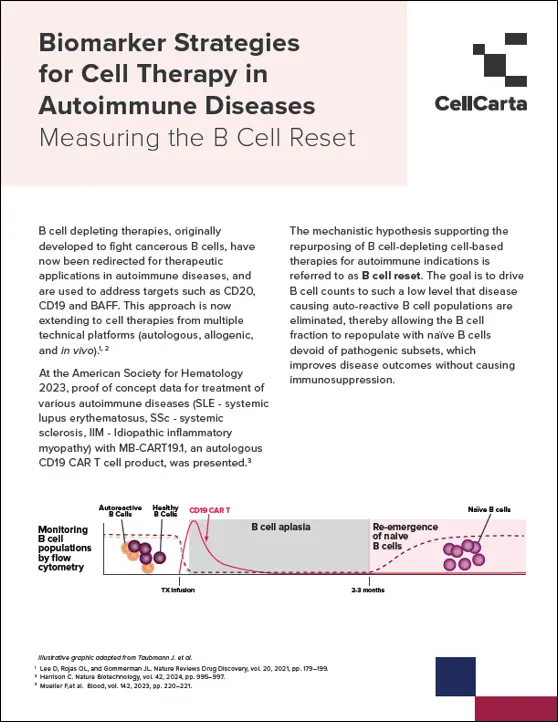
Biomarker Strategies for Cell Therapy in Autoimmune Diseases Measuring the B Cell Reset

Biomarker Strategies for Cell Therapy in Autoimmune Diseases

October 16, 2024

Biomarker Strategies for Cell Therapy in Autoimmune Diseases Measuring the B Cell Reset

Drug Target Engagement with Flow Cytometry Receptor Occupancy Assays

September 17, 2024

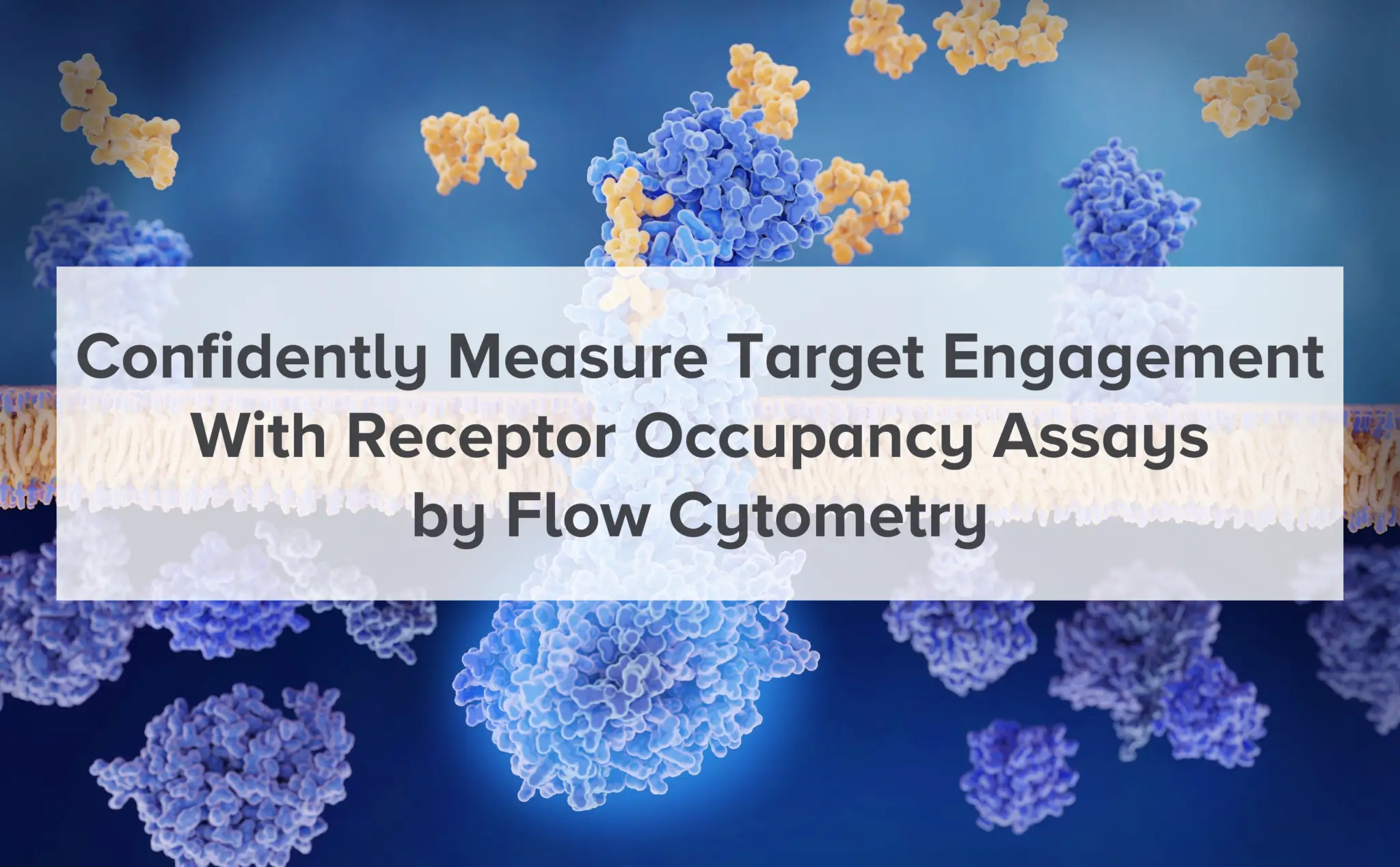
Receptor Occupancy Assays by Flow Cytometry

A crucial step in the drug development process involves optimizing drug-target engagement for your biotherapeutic and gaining valuable pharmacodynamic biomarker data. High-quality receptor occupancy assays (RO assays) are vital tools in this process.

Reliable and accurate results in the design, development, and implementation of receptor occupancy assays can seem to be a tasking prospect, as they are prone to numerous technical and logistical challenges, requiring-

  • expert assay design
  • optimal matrix selection
  • data normalization/reporting
  • and rigorous quality control1, 2

Such challenges can escalate further in difficult development scenarios, such as when the target antigen is expressed at low levels, where there is receptor modulation, or when the therapeutic molecules are bi-specific and bind multiple targets2.

Types and Methodologies

RO assays can be classified into two main types: competitive assays and saturation assays. Competitive assays involve the use of competitive and non-competitive antibodies to the drug, to note that the competitive antibody can also be substituted by an anti-drug antibody. Saturation assays use a competitive antibody and the drug product itself detected by an antibody as a reference point.

Type of Assay Description
Competing vs. non-competing antibodies Competing and non-competing antibodies are added to a sample. Competing antibodies bind to the drug's target site, indicating unbound targets. Non-competing antibodies bind elsewhere on the target, showing total available targets. Alternatively, the competing antibody can be substituted for a anti-drug antibody
Saturation assay Half of the sample is saturated with the drug, mimicking 100% RO, showing the total number of available target sites. The other half remains unsaturated, reflecting drug binding in the patient.
A secondary antibody detects the drug, this can be done using an anti-drug or an anti-Ig antibody, and the ratio of unsaturated to saturated samples reveals the drug's receptor occupancy (RO) level.

Custom RO Strategies for Enhanced Drug Development

CellCarta’s flow-cytometry-based receptor occupancy (RO) assays are designed to overcome these challenges and accelerate your efforts, providing you with the critical information needed to demonstrate target engagement, and gain insight into what degree and how long your biotherapeutic binds its target.

Our RO assays can also be used to complement your pharmacokinetic profiling to provide valuable information on dose selection and frequency of drug infusion. Additionally, RO assays can be validated to support secondary endpoints.

One of our RO strategies starts with the identification of both a competitive and a non-competitive antibody. The competitive antibody will only bind to its target if it is not currently bound by the drug, allowing identification of free receptors, while the non-competitive antibody identifies the total amount of target receptors.

We monitor receptor occupancy only in cell populations of interest by combining target-specific reagents into a flow cytometry panel of phenotypic markers. Competitive and non-competitive antibodies can even be used in the same panel.

When competitive or non-competitive antibodies cannot be identified, or when antibodies to the receptor are not available, we use a saturating vs. non-saturating approach to determine the receptor occupancy or RO.

The strategy involves saturation of half of the sample, mimicking a 100% RO. The other half is not saturated, allowing us to perform a ratio of drug-binding between the two halves to accurately determine the receptor occupancy of the sample.

Pharmacokinetic and Pharmacodynamic Insights

RO assays are integrated with pharmacokinetic (PK) and pharmacodynamic (PD) models to inform dosing strategies and optimize drug efficacy. These assays help determine the relationship between drug concentration and its biological effect, providing critical data for dose selection and frequency of administration. This integration is particularly valuable in clinical trials, where RO assays can serve as pharmacodynamic biomarker measurements to assess drug efficacy.

Choosing the ideal sample matrix for accurate RO Assays

The choice of sample matrix can have a profound effect on the quality of a RO assay. For example, PBMC processing can negatively impact the binding of the drug, resulting in an underestimation of the RO. We also typically test different vacutainers to maximize the stability and precision of the RO measurement.

Our experienced scientists develop and validate different RO strategies to address a variety of drug types and reagent availabilities, drawing on our extensive experience in deploying RO assay strategies in clinical trials.

Challenges and Considerations

Despite their importance, RO assays present several challenges. The development and optimization of these assays are complex and demand high-quality reagents and rigorous controls. Additionally, interpreting the data can be complicated by factors such as receptor internalization and degradation, necessitating a thorough understanding of the underlying biological processes.

Emerging Trends and Technological Breakthroughs in Receptor Occupancy Assays

Advanced technologies and methodologies are continually enhancing the accuracy and reliability of RO assays. Complex RO assays can provide additional insights into receptor internalization and shedding, contributing to a more comprehensive understanding of drug-target interactions. These advancements hold the potential to revolutionize drug development by providing more precise and detailed pharmacodynamic data.

CellCarta exemplifies these advancements with state-of-the-art flow cytometry and custom panels that ensure precise measurements, even in complex scenarios. This accuracy is crucial for informing dose selection and optimizing therapeutic efficacy. Emerging technologies like single-cell RNA sequencing and high-dimensional flow cytometry promise even deeper insights into drug-receptor interactions.

Step Up to the Next Level in Your RO Assay Development and Validation

Enhance the robustness of your RO assay development with our custom-designed panels and expert guidance, ensuring accurate and reliable data that drives informed decision-making in your drug development process.

Expert Insights: Watch the following video from our expert to mastering receptor occupancy assays with Flow Cytometry


About the author:

author photo

Damien Montamat-Sicotte is a Scientific Business Director at CellCarta, specializing in the flow cytometry platform. With a PhD in immunology and post-doctoral expertise from various institutions, Damien has profuse experience in managing the processing and analysis of clinical samples by flow cytometry in an immune monitoring context.

References

  1. Liang M, Schwickart M, Schneider AK, et al. Receptor occupancy assessment by flow cytometry as a pharmacodynamic biomarker in biopharmaceutical development. Cytometry B Clin Cytom 2016;90:117-27.
  2. Hilt E, Sun YS, McCloskey TW, et al. Best practices for optimization and validation of flow cytometry-based receptor occupancy assays. Cytometry B Clin Cytom 2021;100:63-71.

Drug Target Engagement with Flow Cytometry Receptor Occupancy Assays

September 17, 2024

Receptor Occupancy Assays by Flow Cytometry

A crucial step in the drug development process involves optimizing drug-target engagement for your biotherapeutic and gaining valuable pharmacodynamic biomarker data. High-quality receptor occupancy assays (RO assays) are vital tools in this process.

Reliable and accurate results in the design, development, and implementation of receptor occupancy assays can seem to be a tasking prospect, as they are prone to numerous technical and logistical challenges, requiring-

  • expert assay design
  • optimal matrix selection
  • data normalization/reporting
  • and rigorous quality control1, 2

Such challenges can escalate further in difficult development scenarios, such as when the target antigen is expressed at low levels, where there is receptor modulation, or when the therapeutic molecules are bi-specific and bind multiple targets2.

Types and Methodologies

RO assays can be classified into two main types: competitive assays and saturation assays. Competitive assays involve the use of competitive and non-competitive antibodies to the drug, to note that the competitive antibody can also be substituted by an anti-drug antibody. Saturation assays use a competitive antibody and the drug product itself detected by an antibody as a reference point.

Type of Assay Description
Competing vs. non-competing antibodies Competing and non-competing antibodies are added to a sample. Competing antibodies bind to the drug's target site, indicating unbound targets. Non-competing antibodies bind elsewhere on the target, showing total available targets. Alternatively, the competing antibody can be substituted for a anti-drug antibody
Saturation assay Half of the sample is saturated with the drug, mimicking 100% RO, showing the total number of available target sites. The other half remains unsaturated, reflecting drug binding in the patient.
A secondary antibody detects the drug, this can be done using an anti-drug or an anti-Ig antibody, and the ratio of unsaturated to saturated samples reveals the drug's receptor occupancy (RO) level.

Custom RO Strategies for Enhanced Drug Development

CellCarta’s flow-cytometry-based receptor occupancy (RO) assays are designed to overcome these challenges and accelerate your efforts, providing you with the critical information needed to demonstrate target engagement, and gain insight into what degree and how long your biotherapeutic binds its target.

Our RO assays can also be used to complement your pharmacokinetic profiling to provide valuable information on dose selection and frequency of drug infusion. Additionally, RO assays can be validated to support secondary endpoints.

One of our RO strategies starts with the identification of both a competitive and a non-competitive antibody. The competitive antibody will only bind to its target if it is not currently bound by the drug, allowing identification of free receptors, while the non-competitive antibody identifies the total amount of target receptors.

We monitor receptor occupancy only in cell populations of interest by combining target-specific reagents into a flow cytometry panel of phenotypic markers. Competitive and non-competitive antibodies can even be used in the same panel.

When competitive or non-competitive antibodies cannot be identified, or when antibodies to the receptor are not available, we use a saturating vs. non-saturating approach to determine the receptor occupancy or RO.

The strategy involves saturation of half of the sample, mimicking a 100% RO. The other half is not saturated, allowing us to perform a ratio of drug-binding between the two halves to accurately determine the receptor occupancy of the sample.

Pharmacokinetic and Pharmacodynamic Insights

RO assays are integrated with pharmacokinetic (PK) and pharmacodynamic (PD) models to inform dosing strategies and optimize drug efficacy. These assays help determine the relationship between drug concentration and its biological effect, providing critical data for dose selection and frequency of administration. This integration is particularly valuable in clinical trials, where RO assays can serve as pharmacodynamic biomarker measurements to assess drug efficacy.

Choosing the ideal sample matrix for accurate RO Assays

The choice of sample matrix can have a profound effect on the quality of a RO assay. For example, PBMC processing can negatively impact the binding of the drug, resulting in an underestimation of the RO. We also typically test different vacutainers to maximize the stability and precision of the RO measurement.

Our experienced scientists develop and validate different RO strategies to address a variety of drug types and reagent availabilities, drawing on our extensive experience in deploying RO assay strategies in clinical trials.

Challenges and Considerations

Despite their importance, RO assays present several challenges. The development and optimization of these assays are complex and demand high-quality reagents and rigorous controls. Additionally, interpreting the data can be complicated by factors such as receptor internalization and degradation, necessitating a thorough understanding of the underlying biological processes.

Emerging Trends and Technological Breakthroughs in Receptor Occupancy Assays

Advanced technologies and methodologies are continually enhancing the accuracy and reliability of RO assays. Complex RO assays can provide additional insights into receptor internalization and shedding, contributing to a more comprehensive understanding of drug-target interactions. These advancements hold the potential to revolutionize drug development by providing more precise and detailed pharmacodynamic data.

CellCarta exemplifies these advancements with state-of-the-art flow cytometry and custom panels that ensure precise measurements, even in complex scenarios. This accuracy is crucial for informing dose selection and optimizing therapeutic efficacy. Emerging technologies like single-cell RNA sequencing and high-dimensional flow cytometry promise even deeper insights into drug-receptor interactions.

Step Up to the Next Level in Your RO Assay Development and Validation

Enhance the robustness of your RO assay development with our custom-designed panels and expert guidance, ensuring accurate and reliable data that drives informed decision-making in your drug development process.

Expert Insights: Watch the following video from our expert to mastering receptor occupancy assays with Flow Cytometry


About the author:

author photo

Damien Montamat-Sicotte is a Scientific Business Director at CellCarta, specializing in the flow cytometry platform. With a PhD in immunology and post-doctoral expertise from various institutions, Damien has profuse experience in managing the processing and analysis of clinical samples by flow cytometry in an immune monitoring context.

References

  1. Liang M, Schwickart M, Schneider AK, et al. Receptor occupancy assessment by flow cytometry as a pharmacodynamic biomarker in biopharmaceutical development. Cytometry B Clin Cytom 2016;90:117-27.
  2. Hilt E, Sun YS, McCloskey TW, et al. Best practices for optimization and validation of flow cytometry-based receptor occupancy assays. Cytometry B Clin Cytom 2021;100:63-71.

How cell therapy developers are future-proofing clinical monitoring of B-cell aplasia

July 11, 2024

As cell therapies become more complex, the pressures facing clinical developers are rising. A greater number and diversity of cell therapy products are progressing to clinical stages, all requiring comprehensive, accurate, and yet rapid characterization to ensure that the journey from bench to bedside is as fast, safe, and effective as possible.

To accommodate this ever-growing number of new cell therapy concepts, clinical developers need robust, adaptive, affordable testing strategies for extensive yet efficient characterization. To realize these, developers are turning to complementary methods and modularized testing, and working to identify the most high-value readouts. Taking such an approach allows developers to assess aspects such as B-cell aplasia, even as the parameters monitored in B-cell populations expand as cell therapies address new indications.

Our new Cell Therapy Trend Report reveals how clinical testing is adapting to the rapidly changing cell therapy landscape. Download the report now to learn more and explore several broad trends to be aware of within the space, spanning the testing areas of HLA typing, cytokine profiling, cell enumeration and vector copy number determination, single-cell analytics, and B-cell aplasia.

Bringing complementary testing to B-cell aplasia

We anticipate that B-cell monitoring will soon become a standard part of clinical testing programs for new cell therapies and indications. Because of this, identifying the most efficient, effective, and appropriate monitoring approaches is of undeniable value.

Complementarity in particular offers huge promise here. By leveraging the synergies and capabilities of different established testing methods, developers can evaluate B-cell therapies more comprehensively to paint a detailed picture of how cell therapies act against B-cells. Such an approach could help to identify differences in body tissues, discriminate between on- and off-tumor activity, and shed light on off-tumor effects (such as the depletion of healthy B cells).

Additionally, as cell therapies increasingly address new indications and therapeutic areas — including autoimmune diseases and solid tumors — complementary assays can adapt to address a wider array of parameters, creating exciting new opportunities for in-depth analysis. Developers will also be able to mine the knowledge gleaned from previous testing to inform the design of new adaptive assays, enhance their clinical testing, and improve specificity.

B-cell aplasia: Understanding its role in cell therapy clinical testing

As well as improving our understanding of how cell therapies act against B cells, the fast, unambiguous enumeration of B cell populations and their depletion can…

  • Inform study inclusion
  • Help reveal the mechanisms behind autoreactivity
  • Support the development of new therapies
  • Potentially serve complementary purposes in the assessment of therapeutic efficacy and disease progression

Complementarity and other approaches to optimize and future-proof the clinical characterization of novel cell therapies, are discussed in our Cell Therapy Trend Report. Download the report now, or contact our team to speak to an expert about your cell therapy clinical testing needs.


About the author

author photo

Liesbet Vervoort is a Group Lead Program Management at CellCarta. With a PhD in immune-oncology and expertise as an operational lab lead and hematopathology program lead, Liesbet has profuse experience in aligning and translating customers’ needs to clinical trial implementation.

How cell therapy developers are future-proofing clinical monitoring of B-cell aplasia

July 11, 2024

As cell therapies become more complex, the pressures facing clinical developers are rising. A greater number and diversity of cell therapy products are progressing to clinical stages, all requiring comprehensive, accurate, and yet rapid characterization to ensure that the journey from bench to bedside is as fast, safe, and effective as possible.

To accommodate this ever-growing number of new cell therapy concepts, clinical developers need robust, adaptive, affordable testing strategies for extensive yet efficient characterization. To realize these, developers are turning to complementary methods and modularized testing, and working to identify the most high-value readouts. Taking such an approach allows developers to assess aspects such as B-cell aplasia, even as the parameters monitored in B-cell populations expand as cell therapies address new indications.

Our new Cell Therapy Trend Report reveals how clinical testing is adapting to the rapidly changing cell therapy landscape. Download the report now to learn more and explore several broad trends to be aware of within the space, spanning the testing areas of HLA typing, cytokine profiling, cell enumeration and vector copy number determination, single-cell analytics, and B-cell aplasia.

Bringing complementary testing to B-cell aplasia

We anticipate that B-cell monitoring will soon become a standard part of clinical testing programs for new cell therapies and indications. Because of this, identifying the most efficient, effective, and appropriate monitoring approaches is of undeniable value.

Complementarity in particular offers huge promise here. By leveraging the synergies and capabilities of different established testing methods, developers can evaluate B-cell therapies more comprehensively to paint a detailed picture of how cell therapies act against B-cells. Such an approach could help to identify differences in body tissues, discriminate between on- and off-tumor activity, and shed light on off-tumor effects (such as the depletion of healthy B cells).

Additionally, as cell therapies increasingly address new indications and therapeutic areas — including autoimmune diseases and solid tumors — complementary assays can adapt to address a wider array of parameters, creating exciting new opportunities for in-depth analysis. Developers will also be able to mine the knowledge gleaned from previous testing to inform the design of new adaptive assays, enhance their clinical testing, and improve specificity.

B-cell aplasia: Understanding its role in cell therapy clinical testing

As well as improving our understanding of how cell therapies act against B cells, the fast, unambiguous enumeration of B cell populations and their depletion can…

  • Inform study inclusion
  • Help reveal the mechanisms behind autoreactivity
  • Support the development of new therapies
  • Potentially serve complementary purposes in the assessment of therapeutic efficacy and disease progression

Complementarity and other approaches to optimize and future-proof the clinical characterization of novel cell therapies, are discussed in our Cell Therapy Trend Report. Download the report now, or contact our team to speak to an expert about your cell therapy clinical testing needs.


About the author

author photo

Liesbet Vervoort is a Group Lead Program Management at CellCarta. With a PhD in immune-oncology and expertise as an operational lab lead and hematopathology program lead, Liesbet has profuse experience in aligning and translating customers’ needs to clinical trial implementation.

How HLA Typing Improves Immunotherapy Precision

December 6, 2023

How can HLA typing drive better immunotherapy development and selection

Immunotherapies hold enormous potential for cancer treatment, but understanding tumor–immune interactions is needed to successfully develop new therapies and identify suitable patients.

Human leucocyte antigen (HLA) typing gives deeper insights into the relationship between the immune system and cancer cells, helping scientists improve immunotherapy administration precision and develop new immunotherapeutic approaches. But what is it?

A deeper dive into HLA typing

HLAs are a combination of different single nucleotide polymorphisms (SNPs) across multiple exons. The HLA system helps the immune system distinguish between self and non-self cells by presenting antigens derived from pathogens, cancer cells, or other foreign entities to immune cells, triggering a response. HLAs are therefore pivotal in cancer immunotherapy — cancer-specific antigens are presented on HLA molecules, which T cells recognize, attack, and kill.

HLA allele and antigen composition varies significantly between individuals, however, so determining their HLA profile — HLA typing — is essential to understand disease onset, progression, and treatment.

How is HLA typing performed?

Most researchers use Sanger sequencing for HLA typing, which detects SNPs. But since both copies of the genes are sequenced together in one reaction, researchers struggle to identify whether the SNPs come from the same (cis) or the opposite (trans) chromosome, making establishing the correct HLA type difficult. Next-generation sequencing (NGS) overcomes cis/trans polymorphism ambiguities by independently identifying and amplifying individual DNA fragments for more accurate typing.

HLA typing in action

Three recent publications highlight the impact of HLA profiling in immunotherapy development:

1.    Immunotherapy in oncology

HLA type affects an individual’s susceptibility to infections or autoimmune diseases, or their oncological immunotherapy response. A 2019 publication discussed how understanding a patient’s HLA profile is vital to effectively tailor immunotherapeutic treatments. Particularly, researchers should match the HLA types of cancer patients with potential donor HLA types to improve the success of treatments like adoptive cell therapy or stem cell therapy.

HLA typing is essential in CAR T-cell therapy, where it helps in the engineering of T cells to target tumor antigens, expanding immunotherapy applications beyond standard treatments.

2.    Precision medicine

HLA genotyping can help researchers to predict the survival of HLA alleles among cancers, based on an underlying cytolytic activity (CYT) mechanism. A recent study showed that a strong HLA allele, in combination with a high tumor mutation burden, could stimulate intensive immune CYT and lead to extended HLA survival. As HLA profiles differ between patients, typing can therefore support oncologists in selecting the most appropriate immunotherapy for an individual.

3.    Viral associated cancer

The 2019 article also highlighted that carcinogenic viruses activate genes that allow cancer cells to escape immune surveillance and proliferate uncontrolled. A patient’s HLA profile plays a significant role in these immune escape mechanisms, leading to the initiation of such cancers and influencing immunotherapy response. Understanding HLA profiles can therefore help effective cancer vaccine design, improving patient outcomes.

Immunotherapy is a promising approach to treat cancers — and HLA typing is key to unlocking its potential. With HLA typing, researchers can better tailor therapies, select the right treatment, and design more effective cancer vaccines.

NGS is critical to getting clearer results from your HLA typing.

Future of HLA Typing: Artificial Intelligence and Predictive Algorithms

As HLA typing evolves, the integration of AI and machine learning is becoming key to advancing immunotherapy.

These technologies are being used to develop predictive models that combine HLA typing with tumor mutational burden (TMB), and other biomarkers, to forecast patient responses to immune checkpoint inhibitors.

This multidisciplinary approach is expected to revolutionize the personalization of cancer treatments.

At CellCarta we developed our own data analysis pipeline to evaluate immune checkpoint biomarkers by looking at RNA sequencing data.

Discover CellCarta’s genomics services.


About the author: 

Author photo

Nathalie Bernard (PhD) is the scientific business director for the Genomics Services unit within CellCarta. Her background is in molecular biology, and she has many years of experience in PCR and sequencing, technologies used to discover or identify DNA and RNA biomarkers of clinical utility. At CellCarta, Nathalie is using her expertise to guide our customers in finding the best solution to their genomic questions.

How HLA Typing Improves Immunotherapy Precision

December 6, 2023

How can HLA typing drive better immunotherapy development and selection

Immunotherapies hold enormous potential for cancer treatment, but understanding tumor–immune interactions is needed to successfully develop new therapies and identify suitable patients.

Human leucocyte antigen (HLA) typing gives deeper insights into the relationship between the immune system and cancer cells, helping scientists improve immunotherapy administration precision and develop new immunotherapeutic approaches. But what is it?

A deeper dive into HLA typing

HLAs are a combination of different single nucleotide polymorphisms (SNPs) across multiple exons. The HLA system helps the immune system distinguish between self and non-self cells by presenting antigens derived from pathogens, cancer cells, or other foreign entities to immune cells, triggering a response. HLAs are therefore pivotal in cancer immunotherapy — cancer-specific antigens are presented on HLA molecules, which T cells recognize, attack, and kill.

HLA allele and antigen composition varies significantly between individuals, however, so determining their HLA profile — HLA typing — is essential to understand disease onset, progression, and treatment.

How is HLA typing performed?

Most researchers use Sanger sequencing for HLA typing, which detects SNPs. But since both copies of the genes are sequenced together in one reaction, researchers struggle to identify whether the SNPs come from the same (cis) or the opposite (trans) chromosome, making establishing the correct HLA type difficult. Next-generation sequencing (NGS) overcomes cis/trans polymorphism ambiguities by independently identifying and amplifying individual DNA fragments for more accurate typing.

HLA typing in action

Three recent publications highlight the impact of HLA profiling in immunotherapy development:

1.    Immunotherapy in oncology

HLA type affects an individual’s susceptibility to infections or autoimmune diseases, or their oncological immunotherapy response. A 2019 publication discussed how understanding a patient’s HLA profile is vital to effectively tailor immunotherapeutic treatments. Particularly, researchers should match the HLA types of cancer patients with potential donor HLA types to improve the success of treatments like adoptive cell therapy or stem cell therapy.

HLA typing is essential in CAR T-cell therapy, where it helps in the engineering of T cells to target tumor antigens, expanding immunotherapy applications beyond standard treatments.

2.    Precision medicine

HLA genotyping can help researchers to predict the survival of HLA alleles among cancers, based on an underlying cytolytic activity (CYT) mechanism. A recent study showed that a strong HLA allele, in combination with a high tumor mutation burden, could stimulate intensive immune CYT and lead to extended HLA survival. As HLA profiles differ between patients, typing can therefore support oncologists in selecting the most appropriate immunotherapy for an individual.

3.    Viral associated cancer

The 2019 article also highlighted that carcinogenic viruses activate genes that allow cancer cells to escape immune surveillance and proliferate uncontrolled. A patient’s HLA profile plays a significant role in these immune escape mechanisms, leading to the initiation of such cancers and influencing immunotherapy response. Understanding HLA profiles can therefore help effective cancer vaccine design, improving patient outcomes.

Immunotherapy is a promising approach to treat cancers — and HLA typing is key to unlocking its potential. With HLA typing, researchers can better tailor therapies, select the right treatment, and design more effective cancer vaccines.

NGS is critical to getting clearer results from your HLA typing.

Future of HLA Typing: Artificial Intelligence and Predictive Algorithms

As HLA typing evolves, the integration of AI and machine learning is becoming key to advancing immunotherapy.

These technologies are being used to develop predictive models that combine HLA typing with tumor mutational burden (TMB), and other biomarkers, to forecast patient responses to immune checkpoint inhibitors.

This multidisciplinary approach is expected to revolutionize the personalization of cancer treatments.

At CellCarta we developed our own data analysis pipeline to evaluate immune checkpoint biomarkers by looking at RNA sequencing data.

Discover CellCarta’s genomics services.


About the author: 

Author photo

Nathalie Bernard (PhD) is the scientific business director for the Genomics Services unit within CellCarta. Her background is in molecular biology, and she has many years of experience in PCR and sequencing, technologies used to discover or identify DNA and RNA biomarkers of clinical utility. At CellCarta, Nathalie is using her expertise to guide our customers in finding the best solution to their genomic questions.

Multiplexed Mass Spec for Immunotherapy Protein Measurement

October 13, 2023

CellCarta’s poster presents a quantitative mass spectrometry workflow designed for the highly multiplexed measurement of immunomodulatory proteins to support immunotherapy clinical trials.

The workflow utilizes immuno-MRM assay panels to quantify up to 113 proteins with high sensitivity and precision, providing valuable data on immune-related biomarkers. This approach enhances the understanding of drug responses and mechanisms of action, crucial for developing new immunotherapies and combination treatments.

The method involves the use of stable-isotope-labeled peptides and immunoaffinity capture, followed by LC-MRM analysis for accurate protein quantification.

Contact us  to gain insights on advanced proteomics solutions.

View the full poster:

A Quantitative Mass Spectrometry Workflow for Highly Multiplexed Measurement of Immunomodulatory Proteins to Support Immunotherapy Clinical Trials

As presented at SITC 2023 AND EORTC 2023

Multiplexed Mass Spec for Immunotherapy Protein Measurement

October 13, 2023

CellCarta’s poster presents a quantitative mass spectrometry workflow designed for the highly multiplexed measurement of immunomodulatory proteins to support immunotherapy clinical trials.

The workflow utilizes immuno-MRM assay panels to quantify up to 113 proteins with high sensitivity and precision, providing valuable data on immune-related biomarkers. This approach enhances the understanding of drug responses and mechanisms of action, crucial for developing new immunotherapies and combination treatments.

The method involves the use of stable-isotope-labeled peptides and immunoaffinity capture, followed by LC-MRM analysis for accurate protein quantification.

Contact us  to gain insights on advanced proteomics solutions.

View the full poster:

A Quantitative Mass Spectrometry Workflow for Highly Multiplexed Measurement of Immunomodulatory Proteins to Support Immunotherapy Clinical Trials

As presented at SITC 2023 AND EORTC 2023