CellCarta Lake Forest | Rapid Clinical Testing

April 14, 2026
We know meeting critical deadlines is key. After developing two new 6-plex mIF assays, we were able to deliver 9,000+ datapoints in <6 weeks, ahead of submission deadlines.

External milestones place significant pressure on early-stage and translational programs.

That was the case during a rheumatoid arthritis study conducted for a biotech company at CellCarta’s Lake Forest, California laboratory. The project required the rapid development of two new multiplex immunofluorescence (mIF) assays and their transition into clinical sample testing within a compressed timeline tied to an upcoming conference submission deadline.

Here, we highlight how the Lake Forest team combined rapid assay validation with coordinated clinical testing to deliver ahead of the sponsor’s deadline (Figure 1).

87 samples – both 6-plex mIF assays completed in 6 weeks and 9,000 data points delivered ahead of schedule.
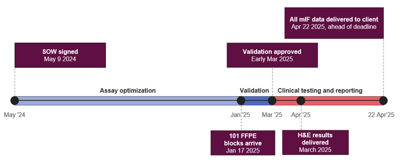
Figure 1: Key milestones of the project at the Lake Forest laboratory. With coordination across teams, the lab was able to move from contract signing to conference-ready data in under 12 months, delivering to the client ahead of schedule.

Optimizing Two New mIF Assays for Rheumatoid Arthritis

The initial phase of the project focused on optimizing two six-plex multiplex immunofluorescence assays on the Akoya platform for use in rheumatoid arthritis tissue samples, a new indication for the Lake Forest Laboratory.

Optimization was carried out between May and December 2024, followed by validation staining and digital image analysis algorithm development in January and February 2025.

Due to the indication, algorithm development proved to be particularly complex, but through careful prioritization and close collaboration between the sponsor, pathology, image analysis scientists, and assay development scientists, the team managed to make quick progress to meet the required deadline.

During this phase, the team also created custom Hematoxylin and Eosin (H&E) evaluation forms for rheumatoid arthritis. These forms were developed to support both assay validation and future clinical sample review, enabling the same tailored pathology evaluation process to be applied across both stages of the project.

Coordinating Development and Clinical Execution

The 101 clinical FFPE block samples arrived at the Lake Forest laboratory on January 17, 2025, while assay validation was being finalized.

To keep the project aligned with the sponsor’s timeline for data delivery by the end of April, the team created a detailed plan for each department to execute as soon as the sponsor approved the full validation package. This close collaboration across assay development, laboratory operations, pathology, digital pathology, and data teams ensured efficient tissue sectioning, staining, and analysis workflows. With this in place, the Lake Forest team was able to:

  • Freshly section all FFPE blocks to retain antigenicity, which is critical in mIF testing.
  • Deliver H&E results in March, allowing the sponsor to quickly review the samples and select those that would proceed to multiplex immunofluorescence analysis.
  • Complete testing and analysis of 87 sponsor-selected samples for both 6-plex mIF assays in just six weeks. All multiplex data, totaling over 9,000 data points (87 samples, each with 105 reported fields), was sent by April 22, ahead of the sponsor’s submission deadline.
  • Deliver the results and associated images in ready-to-use formats ahead of the deadline, enabling the sponsor to review the findings and incorporate the data directly into their conference presentation.

Maintaining Momentum When Timelines Are Fixed

Scientific and translational programs often operate against immovable deadlines. When that happens, sponsors need partners who can move quickly without compromising scientific rigor.

At Lake Forest, close coordination between assay development, pathology, and clinical operations allowed complex assay work and large-scale sample testing to proceed in parallel, helping the sponsor deliver critical data in time for their submission milestone.

Looking for a scientific partner for your next study? Connect with our team to find out how we can support your program.

About CellCarta’s California, Lake Forest Laboratory

For over 20 years, CellCarta’s CLIA and CAP-accredited Lake Forest laboratory has supported pharmaceutical and biotechnology sponsors across preclinical, discovery, and clinical development programs. Key capabilities include:

  • Multiplex immunohistochemistry and immunofluorescence expertise
  • Integrated digital pathology and spatial analysis workflows
  • In-house tissue biobank access
  • Rapid turnaround times designed for early-stage decision-making

Since its opening, the Lake Forest team has worked with over 230 sponsors on over 2,400 unique projects. Operating independently and capable of participating in multi-site joint studies, Lake Forest delivers the speed and flexibility required to advance early-stage programs with confidence.

Find out more about our Lake Forest lab and how it can support your early-stage programs.

About the Author:
author photo

Andrea Reigel is the Director of US Project Management at CellCarta, leading teams across Lake Forest, CA and Naperville, IL in the delivery of complex clinical trial projects. With a B.S. in Biology, an MBA, and a PMP certification, she brings a mix of scientific knowledge, project management expertise, and strategic leadership to every engagement. At CellCarta, she guides major client partnerships, builds and develops operational teams, and drives process improvements that elevate customer service and project execution.
CellCarta Lake Forest | Rapid Clinical Testing

April 14, 2026

We know meeting critical deadlines is key. After developing two new 6-plex mIF assays, we were able to deliver 9,000+ datapoints in <6 weeks, ahead of submission deadlines.

External milestones place significant pressure on early-stage and translational programs.

That was the case during a rheumatoid arthritis study conducted for a biotech company at CellCarta’s Lake Forest, California laboratory. The project required the rapid development of two new multiplex immunofluorescence (mIF) assays and their transition into clinical sample testing within a compressed timeline tied to an upcoming conference submission deadline.

Here, we highlight how the Lake Forest team combined rapid assay validation with coordinated clinical testing to deliver ahead of the sponsor’s deadline (Figure 1).

87 samples – both 6-plex mIF assays completed in 6 weeks and 9,000 data points delivered ahead of schedule.

Figure 1: Key milestones of the project at the Lake Forest laboratory. With coordination across teams, the lab was able to move from contract signing to conference-ready data in under 12 months, delivering to the client ahead of schedule.

Optimizing Two New mIF Assays for Rheumatoid Arthritis

The initial phase of the project focused on optimizing two six-plex multiplex immunofluorescence assays on the Akoya platform for use in rheumatoid arthritis tissue samples, a new indication for the Lake Forest Laboratory.

Optimization was carried out between May and December 2024, followed by validation staining and digital image analysis algorithm development in January and February 2025.

Due to the indication, algorithm development proved to be particularly complex, but through careful prioritization and close collaboration between the sponsor, pathology, image analysis scientists, and assay development scientists, the team managed to make quick progress to meet the required deadline.

During this phase, the team also created custom Hematoxylin and Eosin (H&E) evaluation forms for rheumatoid arthritis. These forms were developed to support both assay validation and future clinical sample review, enabling the same tailored pathology evaluation process to be applied across both stages of the project.

Coordinating Development and Clinical Execution

The 101 clinical FFPE block samples arrived at the Lake Forest laboratory on January 17, 2025, while assay validation was being finalized.

To keep the project aligned with the sponsor’s timeline for data delivery by the end of April, the team created a detailed plan for each department to execute as soon as the sponsor approved the full validation package. This close collaboration across assay development, laboratory operations, pathology, digital pathology, and data teams ensured efficient tissue sectioning, staining, and analysis workflows. With this in place, the Lake Forest team was able to:

  • Freshly section all FFPE blocks to retain antigenicity, which is critical in mIF testing.
  • Deliver H&E results in March, allowing the sponsor to quickly review the samples and select those that would proceed to multiplex immunofluorescence analysis.
  • Complete testing and analysis of 87 sponsor-selected samples for both 6-plex mIF assays in just six weeks. All multiplex data, totaling over 9,000 data points (87 samples, each with 105 reported fields), was sent by April 22, ahead of the sponsor’s submission deadline.
  • Deliver the results and associated images in ready-to-use formats ahead of the deadline, enabling the sponsor to review the findings and incorporate the data directly into their conference presentation.

Maintaining Momentum When Timelines Are Fixed

Scientific and translational programs often operate against immovable deadlines. When that happens, sponsors need partners who can move quickly without compromising scientific rigor.

At Lake Forest, close coordination between assay development, pathology, and clinical operations allowed complex assay work and large-scale sample testing to proceed in parallel, helping the sponsor deliver critical data in time for their submission milestone.

Looking for a scientific partner for your next study? Connect with our team to find out how we can support your program.

About CellCarta’s California, Lake Forest Laboratory

For over 20 years, CellCarta’s CLIA and CAP-accredited Lake Forest laboratory has supported pharmaceutical and biotechnology sponsors across preclinical, discovery, and clinical development programs. Key capabilities include:

  • Multiplex immunohistochemistry and immunofluorescence expertise
  • Integrated digital pathology and spatial analysis workflows
  • In-house tissue biobank access
  • Rapid turnaround times designed for early-stage decision-making

Since its opening, the Lake Forest team has worked with over 230 sponsors on over 2,400 unique projects. Operating independently and capable of participating in multi-site joint studies, Lake Forest delivers the speed and flexibility required to advance early-stage programs with confidence.

Find out more about our Lake Forest lab and how it can support your early-stage programs.

About the Author:

author photo

Andrea Reigel is the Director of US Project Management at CellCarta, leading teams across Lake Forest, CA and Naperville, IL in the delivery of complex clinical trial projects. With a B.S. in Biology, an MBA, and a PMP certification, she brings a mix of scientific knowledge, project management expertise, and strategic leadership to every engagement. At CellCarta, she guides major client partnerships, builds and develops operational teams, and drives process improvements that elevate customer service and project execution.

Optimizing your in situ hybridization for success

August 30, 2024

What is In Situ Hybridization (ISH)?

in situ hybridization (ISH) is a technique that enables the localization of specific DNA or RNA sequences within tissue sections and cytology samples using labeled complementary strands. The target gene can be visualized through brightfield or fluorescence microscopy. Compared to immunohistochemistry (IHC), which broadly localizes proteins within tissues, ISH offers greater sensitivity, allowing researchers to understand the organization and function of genes in tissue sections. ISH not only identifies cells positive for a specific DNA/RNA sequence but also quantifies the level of positivity within individual cells.

How can you benefit from combining ISH and IHC assays

ISH is a powerful tool in both preclinical research and clinical trials, offering critical insights. It can be combined with IHC assays to provide unique perspectives on cellular and molecular mechanisms within tissues.

Key applications include:

  • Relationship Between Targets: Dual assays on unique but related targets reveal their interrelationships.
  • Protein Localization: Identify the location of a target protein within tissue and the originating cells.
  • Gene Regulation Insight: Combining ISH and IHC on the same target provides insights into gene regulation mechanisms.
  • Tumor Region Analysis: Use IHC cytokeratin markers with ISH assays to highlight gene regulation in tumor regions.
  • Protein Degradation Insights: Dual assays offer valuable insights into protein degradation processes.

Maximizing your in situ hybridization assays

Successful ISH assays depend on meticulous sample preparation to minimize RNA degradation in tissue.

  • Tissue Fixation: Fix tissues promptly in 10% Neutral Buffered Formalin (NBF) at 20x the tissue sample volume, then transfer to 70% ethanol (EtOH).
  • RNA/DNA Preservation: Cut fresh sections under RNase/DNase-free conditions onto charged slides for better adhesion, and store at a constant 4°C or at room temperature.
  •  Baking conditions: Bake slides for 30-120 min at 60°C for better adhesion.

Another crucial aspect for a successful ISH assay is optimization. Each unique combination of species, tissue type, and gene target must be optimized to hone in on the ideal conditions for detecting and visualizing the target. Pre-existing IHC data can guide the optimization process.

Additionally, digital whole slide imaging settings must be tailored to each sample set, accounting for tissue type differences, stain intensity, and autofluorescence.

Consistency in applying the ISH protocol across samples is key. Automated technologies, like the Leica BondRX and Ventana Discovery Ultra, ensure precise and consistent application, minimizing human error.

Leveraging AI-Powered Data for your ISH Assays

The integration of AI-powered algorithms with digital whole slide ISH images enhances the analysis, generating data and insights beyond human capability. However, several key steps ensure successful ISH assays.

Our AI-powered algorithms ensure optimal nuclear segmentation for the most reliable results.

Rest assured that our in-house team of pathologists work in proximity with our imaging specialists to ensure you get optimized data from your ISH assays, that signal is neither undercounted nor overestimated.

By leveraging cutting-edge ISH technologies, CellCarta ensures comprehensive and high-quality results for your research and diagnostic needs.

Contact us to get your project started.


Our expert:

author photo

Sofie Van Rossom (PhD) is a scientific matter expert for ISH technologies within CellCarta. With a background is Biochemistry & Biotechnology, and many years of experience in the development and validation of new (F)ISH assays  to identify DNA and RNA biomarkers of clinical utility, Sofie uses her expertise to guide customers in the validation of their (F)ISH assays.

Optimizing your in situ hybridization for success

August 30, 2024

What is In Situ Hybridization (ISH)?

in situ hybridization (ISH) is a technique that enables the localization of specific DNA or RNA sequences within tissue sections and cytology samples using labeled complementary strands. The target gene can be visualized through brightfield or fluorescence microscopy. Compared to immunohistochemistry (IHC), which broadly localizes proteins within tissues, ISH offers greater sensitivity, allowing researchers to understand the organization and function of genes in tissue sections. ISH not only identifies cells positive for a specific DNA/RNA sequence but also quantifies the level of positivity within individual cells.

How can you benefit from combining ISH and IHC assays

ISH is a powerful tool in both preclinical research and clinical trials, offering critical insights. It can be combined with IHC assays to provide unique perspectives on cellular and molecular mechanisms within tissues.

Key applications include:

  • Relationship Between Targets: Dual assays on unique but related targets reveal their interrelationships.
  • Protein Localization: Identify the location of a target protein within tissue and the originating cells.
  • Gene Regulation Insight: Combining ISH and IHC on the same target provides insights into gene regulation mechanisms.
  • Tumor Region Analysis: Use IHC cytokeratin markers with ISH assays to highlight gene regulation in tumor regions.
  • Protein Degradation Insights: Dual assays offer valuable insights into protein degradation processes.

Maximizing your in situ hybridization assays

Successful ISH assays depend on meticulous sample preparation to minimize RNA degradation in tissue.

  • Tissue Fixation: Fix tissues promptly in 10% Neutral Buffered Formalin (NBF) at 20x the tissue sample volume, then transfer to 70% ethanol (EtOH).
  • RNA/DNA Preservation: Cut fresh sections under RNase/DNase-free conditions onto charged slides for better adhesion, and store at a constant 4°C or at room temperature.
  •  Baking conditions: Bake slides for 30-120 min at 60°C for better adhesion.

Another crucial aspect for a successful ISH assay is optimization. Each unique combination of species, tissue type, and gene target must be optimized to hone in on the ideal conditions for detecting and visualizing the target. Pre-existing IHC data can guide the optimization process.

Additionally, digital whole slide imaging settings must be tailored to each sample set, accounting for tissue type differences, stain intensity, and autofluorescence.

Consistency in applying the ISH protocol across samples is key. Automated technologies, like the Leica BondRX and Ventana Discovery Ultra, ensure precise and consistent application, minimizing human error.

Leveraging AI-Powered Data for your ISH Assays

The integration of AI-powered algorithms with digital whole slide ISH images enhances the analysis, generating data and insights beyond human capability. However, several key steps ensure successful ISH assays.

Our AI-powered algorithms ensure optimal nuclear segmentation for the most reliable results.

Rest assured that our in-house team of pathologists work in proximity with our imaging specialists to ensure you get optimized data from your ISH assays, that signal is neither undercounted nor overestimated.

By leveraging cutting-edge ISH technologies, CellCarta ensures comprehensive and high-quality results for your research and diagnostic needs.

Contact us to get your project started.


Our expert:

author photo

Sofie Van Rossom (PhD) is a scientific matter expert for ISH technologies within CellCarta. With a background is Biochemistry & Biotechnology, and many years of experience in the development and validation of new (F)ISH assays  to identify DNA and RNA biomarkers of clinical utility, Sofie uses her expertise to guide customers in the validation of their (F)ISH assays.

Comprehensive Histology and Pathology Services by CellCarta

November 22, 2022

Explore Our Advanced Histopathology Laboratories!

As a leading contract research organization (CRO), we offer a wide array of histopathology services.

Our histopathology facilities have trained in-house pathologists and multiple platforms to partner with you and provide integrated solutions for your specific clinical trial needs.

We deliver preclinical and clinical histology support to pharmaceutical and biotech sponsors worldwide.

Discover how CellCarta advances histopathology research and services.

We offer testing services for all phases of clinical trials including:

As a CAP/CLIA-certified CRO Lab, we support clinical studies for diagnostic and pharmaceutical partners in preparation for FDA, EU, and other global submissions.

Our digital pathology services and AI powered workflows provide scalable solutions for high-throughput histology testing across disease areas.

Histology CRO and Pathology CRO Services

Building on our comprehensive services, CellCarta’s Histology CRO capabilities extend to custom assay development and validation, supporting a wide range of clinical and research applications. 

Our expert team provides histology support for CROs, offering tailored solutions to meet the specific requirements of each study. This includes detailed consultation on sample preparation, staining protocols, and data interpretation.

We offers both off-the-shelf and customized  routine and advanced digital histology services, with an expert team ensuring reproducibility, scalability, and regulatory readiness.

Advanced Molecular and Digital Pathology Services

In addition to our core services, we provide advanced molecular pathology services, including in situ hybridization (ISH) and fluorescence in situ hybridization (FISH). 

These techniques enable the detection of specific biomarkers and genetic materials within tissue samples, aiding in the identification and characterization of diseases.

Our digital pathology platforms, featuring whole slide imaging and automated image analysis, allows for accurate, reproducible data generation and efficient workflow management.

Collaborations and Software Solutions

We collaborate with industry leaders to enhance our service offerings and stay at the forefront of histopathology innovations. 

Our histopathology software, including advanced AI-driven platforms, streamline data analysis and provide deeper insights into your samples.

This integration of cutting-edge technology ensures that our clients receive the most reliable and insightful data possible, ensuring CellCarta provides top CRO pathology services for your oncology, immunology, and biomarker programs regardless of disease area.

Quality Assurance and Global Compliance

Our commitment to quality goes beyond our technical capabilities. As a CAP/CLIA certified lab, we adhere to stringent quality control measures and global standards. 

This ensures that our data and analyses are robust, reproducible, and ready for regulatory submissions worldwide, including FDA and EU approvals. 

Our comprehensive quality assurance program includes regular proficiency testing and adherence to best practices in laboratory operations.

Why Choose CellCarta?

CellCarta offers unparalleled expertise in histopathology CRO services, making us a trusted partner for pharmaceutical companies and research institutions.

Whether you require IHC CRO services, digital pathology support, or full-service histopathology assay development, our team is equipped to deliver high-quality data.

Our comprehensive range of services, from routine histology to advanced molecular analyses, combined with our commitment to quality and innovation, ensures that we deliver accurate, high-quality data tailored to your specific needs.

Comprehensive Histology and Pathology Services by CellCarta

November 22, 2022

Explore Our Advanced Histopathology Laboratories!

As a leading contract research organization (CRO), we offer a wide array of histopathology services.

Our histopathology facilities have trained in-house pathologists and multiple platforms to partner with you and provide integrated solutions for your specific clinical trial needs.

We deliver preclinical and clinical histology support to pharmaceutical and biotech sponsors worldwide.

Discover how CellCarta advances histopathology research and services.

We offer testing services for all phases of clinical trials including:

As a CAP/CLIA-certified CRO Lab, we support clinical studies for diagnostic and pharmaceutical partners in preparation for FDA, EU, and other global submissions.

Our digital pathology services and AI powered workflows provide scalable solutions for high-throughput histology testing across disease areas.

Histology CRO and Pathology CRO Services

Building on our comprehensive services, CellCarta’s Histology CRO capabilities extend to custom assay development and validation, supporting a wide range of clinical and research applications. 

Our expert team provides histology support for CROs, offering tailored solutions to meet the specific requirements of each study. This includes detailed consultation on sample preparation, staining protocols, and data interpretation.

We offers both off-the-shelf and customized  routine and advanced digital histology services, with an expert team ensuring reproducibility, scalability, and regulatory readiness.

Advanced Molecular and Digital Pathology Services

In addition to our core services, we provide advanced molecular pathology services, including in situ hybridization (ISH) and fluorescence in situ hybridization (FISH). 

These techniques enable the detection of specific biomarkers and genetic materials within tissue samples, aiding in the identification and characterization of diseases.

Our digital pathology platforms, featuring whole slide imaging and automated image analysis, allows for accurate, reproducible data generation and efficient workflow management.

Collaborations and Software Solutions

We collaborate with industry leaders to enhance our service offerings and stay at the forefront of histopathology innovations. 

Our histopathology software, including advanced AI-driven platforms, streamline data analysis and provide deeper insights into your samples.

This integration of cutting-edge technology ensures that our clients receive the most reliable and insightful data possible, ensuring CellCarta provides top CRO pathology services for your oncology, immunology, and biomarker programs regardless of disease area.

Quality Assurance and Global Compliance

Our commitment to quality goes beyond our technical capabilities. As a CAP/CLIA certified lab, we adhere to stringent quality control measures and global standards. 

This ensures that our data and analyses are robust, reproducible, and ready for regulatory submissions worldwide, including FDA and EU approvals. 

Our comprehensive quality assurance program includes regular proficiency testing and adherence to best practices in laboratory operations.

Why Choose CellCarta?

CellCarta offers unparalleled expertise in histopathology CRO services, making us a trusted partner for pharmaceutical companies and research institutions.

Whether you require IHC CRO services, digital pathology support, or full-service histopathology assay development, our team is equipped to deliver high-quality data.

Our comprehensive range of services, from routine histology to advanced molecular analyses, combined with our commitment to quality and innovation, ensures that we deliver accurate, high-quality data tailored to your specific needs.

CellCarta Naperville Lab | Genomic and Histopathology Services

October 12, 2022

Step Into Our Naperville Facility!

In 2022, we proudly announced the grand opening of our Naperville, Chicago location!

Our new facility is only 30 minutes from the Chicago O’Hare airport and encompasses over 3,000m2 (29,283 sq ft) of innovative scientific equipment and highly trained staff.

As part of CellCarta’s global CRO infrastructure, this lab strengthens our clinical trial support capabilities across North America.

Join us for a tour and see how CellCarta is dedicated to advancing scientific research.

At Naperville, we offer testing services for all phases of clinical trials. As a CRO with multiomics capabilities, we support:

Genomics

Our Naperville facility is designed to provide comprehensive testing services across various phases of clinical trials. Our advanced genomic capabilities include-

  • RT-PCR

These techniques are essential for precise genetic analysis and biomarker discovery, aiding in the development of targeted therapies and personalized medicine.

We support both preclinical and clinical studies across oncology and immunology, as well as specialized therapeutic development such as ADC programs.

Histopathology

In addition to our robust genomic services, we offer state-of-the-art histopathology services.

  • Software platforms include: Indica Labs’ HALO®, Visiopharm®,  Akoya’s inForm®, CyteHub®, SoloWeb, and navify® by Roche.

As a CAP/CLIA certified lab, we support clinical studies for diagnostic and pharmaceutical partners in preparation for FDA, EU and other global submissions.

Our histology CRO services are integrated into our multiomic CRO offerings, enabling scalable analysis pipelines for high-volume biomarker testing across platforms and therapeutic areas.

We support both preclinical and clinical studies across oncology, immunology, and more, with specialized support for ADC development programs.

Commitment to Quality and Compliance

As a CAP accredited and CLIA certified laboratory, we adhere to the highest standards of quality and compliance.

Our rigorous quality control measures ensure the reliability and accuracy of our data, supporting clinical studies for regulatory submissions worldwide, including FDA and EU approvals.

Our quality management system (QMS) drives harmonization of specific testing services and related activities and is implemented at all levels of the organization.

Strategic Location and Accessibility

Located just 30 minutes from Chicago O’Hare airport, our Naperville facility is easily accessible for local and international clients. The facility spans over 3,000m² (29,283 sq ft) and is equipped with the latest scientific equipment, operated by a team of highly trained professionals dedicated to advancing scientific research.

Why Choose CellCarta?

CellCarta is committed to delivering high-quality, reliable data through our extensive range of testing services and advanced technologies. Our new Naperville facility exemplifies our dedication to supporting clinical trials and research initiatives with state-of-the-art resources and expert staff.

As a global CRO, our Naperville site provides key clinical trial services in genomics, histopathology, and biomarker programs.

Backed by our expert scientific teams and cutting-edge platforms, we help biopharma clients accelerate drug development with data they can trust.

CellCarta Naperville Lab | Genomic and Histopathology Services

October 12, 2022

Step Into Our Naperville Facility!

In 2022, we proudly announced the grand opening of our Naperville, Chicago location!

Our new facility is only 30 minutes from the Chicago O’Hare airport and encompasses over 3,000m2 (29,283 sq ft) of innovative scientific equipment and highly trained staff.

As part of CellCarta’s global CRO infrastructure, this lab strengthens our clinical trial support capabilities across North America.

Join us for a tour and see how CellCarta is dedicated to advancing scientific research.

At Naperville, we offer testing services for all phases of clinical trials. As a CRO with multiomics capabilities, we support:

Genomics

Our Naperville facility is designed to provide comprehensive testing services across various phases of clinical trials. Our advanced genomic capabilities include-

  • RT-PCR

These techniques are essential for precise genetic analysis and biomarker discovery, aiding in the development of targeted therapies and personalized medicine.

We support both preclinical and clinical studies across oncology and immunology, as well as specialized therapeutic development such as ADC programs.

Histopathology

In addition to our robust genomic services, we offer state-of-the-art histopathology services.

  • Software platforms include: Indica Labs’ HALO®, Visiopharm®,  Akoya’s inForm®, CyteHub®, SoloWeb, and navify® by Roche.

As a CAP/CLIA certified lab, we support clinical studies for diagnostic and pharmaceutical partners in preparation for FDA, EU and other global submissions.

Our histology CRO services are integrated into our multiomic CRO offerings, enabling scalable analysis pipelines for high-volume biomarker testing across platforms and therapeutic areas.

We support both preclinical and clinical studies across oncology, immunology, and more, with specialized support for ADC development programs.

Commitment to Quality and Compliance

As a CAP accredited and CLIA certified laboratory, we adhere to the highest standards of quality and compliance.

Our rigorous quality control measures ensure the reliability and accuracy of our data, supporting clinical studies for regulatory submissions worldwide, including FDA and EU approvals.

Our quality management system (QMS) drives harmonization of specific testing services and related activities and is implemented at all levels of the organization.

Strategic Location and Accessibility

Located just 30 minutes from Chicago O’Hare airport, our Naperville facility is easily accessible for local and international clients. The facility spans over 3,000m² (29,283 sq ft) and is equipped with the latest scientific equipment, operated by a team of highly trained professionals dedicated to advancing scientific research.

Why Choose CellCarta?

CellCarta is committed to delivering high-quality, reliable data through our extensive range of testing services and advanced technologies. Our new Naperville facility exemplifies our dedication to supporting clinical trials and research initiatives with state-of-the-art resources and expert staff.

As a global CRO, our Naperville site provides key clinical trial services in genomics, histopathology, and biomarker programs.

Backed by our expert scientific teams and cutting-edge platforms, we help biopharma clients accelerate drug development with data they can trust.