Powering histopathology with digital pathology solutions and image analysis
Transforming Pathology Practice with Innovative Digital Imaging Solutions
As a first adopter of digital whole slide high-resolution image capture and with nearly two decades of operations, we have resolved and pioneered multiple imaging and image analysis methodologies that are ideal for clinical trial deployment.
All our services are guided by carefully optimized and rigorously documented procedures.
Key services include:
- Pathologist-directed image analysis
- Digital whole-slide brightfield and fluorescent scanning
- Multispectral imaging
- Dedicated team of imaging scientists, image analysts, and application experts, implementing customized and commercial algorithms
Each slide is digitally captured in high-resolution, whole-slide format and retained on our image servers.
The larger part of the evaluation and scoring is performed by our team of trained pathologists and imaging scientists.
Image scanning and analysis for clinical trials are performed in a CLIA-certified, CAP-accredited laboratory.
Whole slide scanning technologies at CellCarta
At CellCarta, we use industry-standard scanning technology, like fluorescent and brightfield scanners from
- 3DHistech
- Akoya
- BioView
- Leica Biosystems (Aperio®)
- Roche/Ventana
- and Zeiss
to enable high throughput, and high-resolution imaging with a fast turnaround time.
Whole slide scanning is a strategic solution for digital image archiving, image analysis, and sharing of staining results.
We can provide whole-slide scanning as part of our end-to-end digital pathology services or as a stand-alone service for your digitization programs.
Contact us to learn more about our services.
Advanced Digital Image Analysis Solutions
Our broad portfolio of image analysis tools includes
- Lunit SCOPE® suite
- Indica Labs’ HALO®
- Visiopharm®,
- Akoya’s inForm®
- CyteHub®
- SoloWeb
- and navify® by Roche
Our team can capture valuable data from tissues including cell phenotypes, densities, and spatial biology running commercially validated assays. In addition to developing custom algorithms that allow us to extract the information that matters for your clinical programs.
Image Analysis Capabilities
- H&E, IHC, IF, single and multiplexed, brightfield and fluorescent image analysis
- Spatial biology measurements and nearest neighbor analysis
- Stereology-based methods for systematic uniform random sampling of ROIs for pathologist scoring e.g. for microvessel density
- Image analysis algorithms developed by our scientists and validated according to CellCarta’s quality standards
Our team of scientists is always available for consultation to help make sense of the expansive datasets generated for your trials.
AI-powered pathology
Our digital pathology workflows are powered by AI.
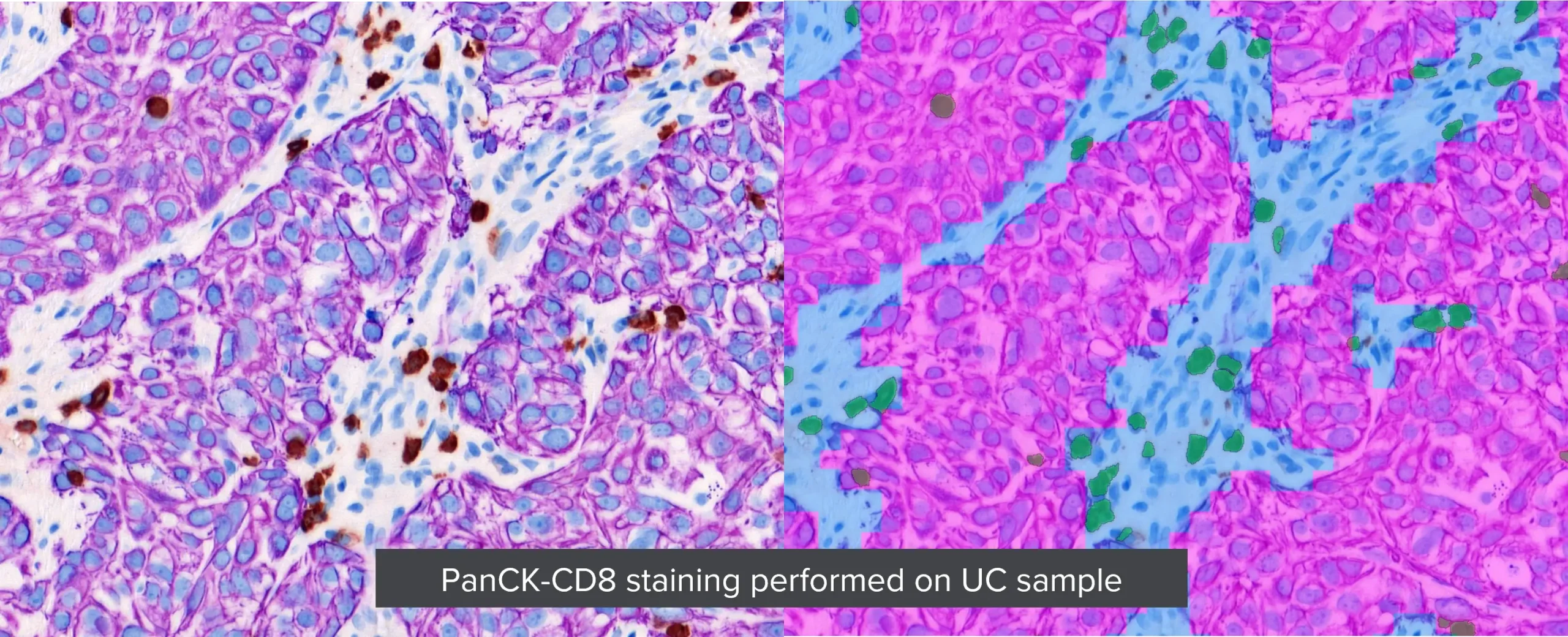
Whether it’s to detect CTCs for nuclear segmentation, tissue classification, or cell phenotyping, AI is embedded in all our image analysis platforms.
We offer our expertise in developing and validating new AI-based algorithms for your studies, or we can collaborate with your preferred digital pathology AI provider to apply their algorithms in your clinical trial assays conducted at CellCarta.