April 16, 2025
Our team developed a comprehensive suite of precision assays to support every stage of antibody-drug conjugate (ADC) development.
From confirming target expression and measuring density, to evaluating drug-target engagement, pharmacokinetics, and safety, CellCarta employs a data-driven, multiplatform approach.
Our solutions include multiplex IF and IHC, flow cytometry, mass spectrometry, RNA sequencing, and spatial biology technologies, ensuring precise target quantification and in-depth tumor microenvironment analysis.
With global facilities, companion diagnostic (CDx) expertise, and full logistics support, CellCarta helps biopharma partners accelerate their ADC programs.

Advance your ADC programs with CellCarta! Contact us today to discover how our precision assays and scientific expertise can drive your clinical success.
April 16, 2025
Our team developed a comprehensive suite of precision assays to support every stage of antibody-drug conjugate (ADC) development.
From confirming target expression and measuring density, to evaluating drug-target engagement, pharmacokinetics, and safety, CellCarta employs a data-driven, multiplatform approach.
Our solutions include multiplex IF and IHC, flow cytometry, mass spectrometry, RNA sequencing, and spatial biology technologies, ensuring precise target quantification and in-depth tumor microenvironment analysis.
With global facilities, companion diagnostic (CDx) expertise, and full logistics support, CellCarta helps biopharma partners accelerate their ADC programs.
Advance your ADC programs with CellCarta! Contact us today to discover how our precision assays and scientific expertise can drive your clinical success.
January 24, 2025

Traditional endpoints in oncology clinical trials, such as progression-free survival (PFS) and overall survival (OS), while definitive, often require extended follow-up periods and substantial patient cohorts. These requirements can significantly extend development timelines and increase costs.
Monitoring tumor burden through measurable residual disease (MRD) assessment — measuring the presence of residual cancer cells during or after treatment — has emerged as a promising alternative, offering an early biomarker of treatment response or relapse that could accelerate clinical development decisions.
This article examines current practices and recent developments in MRD measurement across both blood and solid malignancies.
In hematologic malignancies, MRD assessment has gained significant traction, with the FDA’s Oncologic Drugs Advisory Committee (ODAC) recently recognizing MRD as an accepted endpoint for accelerated approval in multiple myeloma studies. MRD in multiple myeloma is currently measured using a highly specific, sensitive CE-marked next-generation sequencing (NGS)-based test, clonoSEQ.
Traditionally, MRD assessment in blood cancers has relied on bone marrow (BM) extracts. However, obtaining BM extracts is an invasive and painful procedure for patients, and may no longer be necessary. In fact, recent research suggests that less-invasive liquid biopsies could be a suitable alternative.
Mass spectrometry (MS) can detect soluble BCMA, a protein expressed on multiple myeloma cells, and M-protein, an abnormal protein produced by precancerous and cancerous bone marrow cells, in peripheral blood. Notably, MS applied to liquid biopsies has demonstrated greater sensitivity compared to NGS-based testing (clonoSEQ) of bone marrow extracts for M-protein detection.
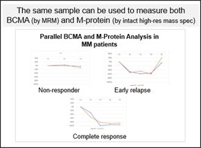
MS offers the distinct advantage of tracking the specific M-protein clone produced by cancer cells and monitoring for additional clones as treatment progresses. This is facilitated by frequent sampling of peripheral blood, allowing for shorter intervals between assessments. Its high analytical range also enables the detection of signal even with reductions exceeding 90% of the original M-Protein amount, providing confidence in the quantification. The data generated by MS complements NGS-based testing and can guide decisions on further testing, including bone marrow biopsies. Importantly, MS can simultaneously measure both M-protein and BCMA from a single sample, making it an efficient tool for assessing tumor burden biomarkers.
Flow cytometry offers another approach for MRD measurement, specifically for detecting the presence of abnormal plasma cells, and can also be used to confirm target expression and identify new targets on abnormal cells. It has several advantages over NGS-based approaches for MRD measurement, including the fact that it is a standalone assay, and that there is no need to normalize to the screening timepoint and may help identify new phenotypes of abnormal cells.
While MRD assessment originated in hematologic malignancies, its application in solid tumors is rapidly evolving.
Measuring MRD in solid tumors is typically done by tracking changes in levels of circulating tumor DNA (ctDNA) — DNA released into the bloodstream by dying tumor cells — using liquid biopsies, which are less invasive than tissue biopsies and enable earlier and more frequent monitoring of tumor burden. Since ctDNA originates from multiple lesions throughout the body, it also provides a more comprehensive view of tumor heterogeneity than single-site tissue biopsies.
Two main approaches have emerged for solid tumor MRD testing:
The FDA recently released new guidance for those planning to use ctDNA assays for curative intent solid tumor drug development, with the guidance dedicating significant focus to ctDNA assay considerations for MRD measurement in particular.
Sequencing technologies offer value beyond MRD monitoring, too. There is a growing focus in oncology trials on understanding whether patients are developing therapeutic resistance, driven by mutations that emerge during treatment.
Advanced sequencing techniques can track these mutational changes throughout treatment, helping to identify resistance pathways and inform therapeutic adaptation. This application is currently most established in solid tumors, though it is also employed, albeit less frequently, in hematologic malignancies.
The ability to monitor mutation drift by sequencing liquid biopsies represents another powerful application of sequencing tools in personalizing cancer treatment, ensuring therapies can be adapted based on a patient’s evolving disease profile.
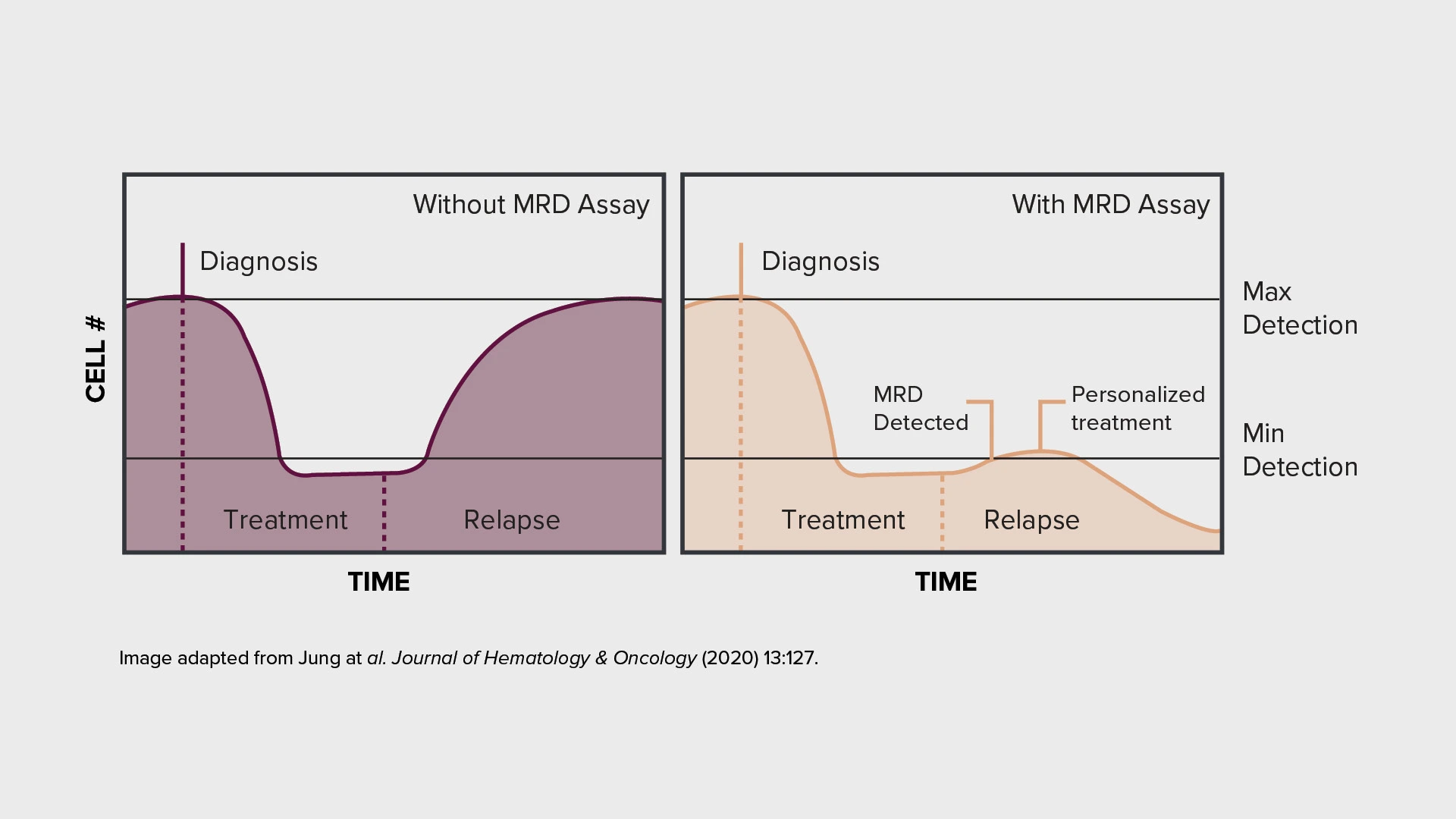
The integration of MRD assessment into clinical trials can offer earlier indication of treatment efficacy and enables more frequent disease monitoring. These capabilities can support more rapid development decisions in oncology trials.
Crucially, the field continues to evolve, with ongoing technological advances improving the sensitivity and reliability of MRD detection. A growing range of validated tools and approaches — from next-generation sequencing to mass spectrometry and flow cytometry — now provides options for accurate MRD measurement across different cancer types and clinical contexts.
Want to find out how MRD measurement could help optimize your clinical studies? Reach out to one of our experts to find out more.
About the authors:

Nathalie Bernard (PhD) is the scientific business director for the Genomic Services unit within CellCarta. Her background is in molecular biology, and she has many years of experience in PCR and sequencing, technologies used to discover or identify DNA and RNA biomarkers of clinical utility. At CellCarta, Nathalie is using her expertise to guide our customers in finding the best solution to their genomic questions.

Dr. Luca Genovesi is the Group Leader of R&D at CellCarta, where he manages a team of scientists in developing mass spectrometry-based assays to quantify biologics and biomarkers in complex matrices for clinical and pre-clinical studies. He has a strong background in Analytical Chemistry, with over 15 years of experience in the regulated pharmaceutical industry, including roles in pharmaceutical companies, CMOs, and CROs. Dr. Genovesi holds an M.Sc. in Organic Chemistry and a Ph.D. in Industrial Biotechnology from Milan University, Italy.
January 24, 2025
Traditional endpoints in oncology clinical trials, such as progression-free survival (PFS) and overall survival (OS), while definitive, often require extended follow-up periods and substantial patient cohorts. These requirements can significantly extend development timelines and increase costs.
Monitoring tumor burden through measurable residual disease (MRD) assessment — measuring the presence of residual cancer cells during or after treatment — has emerged as a promising alternative, offering an early biomarker of treatment response or relapse that could accelerate clinical development decisions.
This article examines current practices and recent developments in MRD measurement across both blood and solid malignancies.
In hematologic malignancies, MRD assessment has gained significant traction, with the FDA’s Oncologic Drugs Advisory Committee (ODAC) recently recognizing MRD as an accepted endpoint for accelerated approval in multiple myeloma studies. MRD in multiple myeloma is currently measured using a highly specific, sensitive CE-marked next-generation sequencing (NGS)-based test, clonoSEQ.
Traditionally, MRD assessment in blood cancers has relied on bone marrow (BM) extracts. However, obtaining BM extracts is an invasive and painful procedure for patients, and may no longer be necessary. In fact, recent research suggests that less-invasive liquid biopsies could be a suitable alternative.
Mass spectrometry (MS) can detect soluble BCMA, a protein expressed on multiple myeloma cells, and M-protein, an abnormal protein produced by precancerous and cancerous bone marrow cells, in peripheral blood. Notably, MS applied to liquid biopsies has demonstrated greater sensitivity compared to NGS-based testing (clonoSEQ) of bone marrow extracts for M-protein detection.
MS offers the distinct advantage of tracking the specific M-protein clone produced by cancer cells and monitoring for additional clones as treatment progresses. This is facilitated by frequent sampling of peripheral blood, allowing for shorter intervals between assessments. Its high analytical range also enables the detection of signal even with reductions exceeding 90% of the original M-Protein amount, providing confidence in the quantification. The data generated by MS complements NGS-based testing and can guide decisions on further testing, including bone marrow biopsies. Importantly, MS can simultaneously measure both M-protein and BCMA from a single sample, making it an efficient tool for assessing tumor burden biomarkers.
Flow cytometry offers another approach for MRD measurement, specifically for detecting the presence of abnormal plasma cells, and can also be used to confirm target expression and identify new targets on abnormal cells. It has several advantages over NGS-based approaches for MRD measurement, including the fact that it is a standalone assay, and that there is no need to normalize to the screening timepoint and may help identify new phenotypes of abnormal cells.
While MRD assessment originated in hematologic malignancies, its application in solid tumors is rapidly evolving.
Measuring MRD in solid tumors is typically done by tracking changes in levels of circulating tumor DNA (ctDNA) — DNA released into the bloodstream by dying tumor cells — using liquid biopsies, which are less invasive than tissue biopsies and enable earlier and more frequent monitoring of tumor burden. Since ctDNA originates from multiple lesions throughout the body, it also provides a more comprehensive view of tumor heterogeneity than single-site tissue biopsies.
Two main approaches have emerged for solid tumor MRD testing:
The FDA recently released new guidance for those planning to use ctDNA assays for curative intent solid tumor drug development, with the guidance dedicating significant focus to ctDNA assay considerations for MRD measurement in particular.
Sequencing technologies offer value beyond MRD monitoring, too. There is a growing focus in oncology trials on understanding whether patients are developing therapeutic resistance, driven by mutations that emerge during treatment.
Advanced sequencing techniques can track these mutational changes throughout treatment, helping to identify resistance pathways and inform therapeutic adaptation. This application is currently most established in solid tumors, though it is also employed, albeit less frequently, in hematologic malignancies.
The ability to monitor mutation drift by sequencing liquid biopsies represents another powerful application of sequencing tools in personalizing cancer treatment, ensuring therapies can be adapted based on a patient’s evolving disease profile.
The integration of MRD assessment into clinical trials can offer earlier indication of treatment efficacy and enables more frequent disease monitoring. These capabilities can support more rapid development decisions in oncology trials.
Crucially, the field continues to evolve, with ongoing technological advances improving the sensitivity and reliability of MRD detection. A growing range of validated tools and approaches — from next-generation sequencing to mass spectrometry and flow cytometry — now provides options for accurate MRD measurement across different cancer types and clinical contexts.
Want to find out how MRD measurement could help optimize your clinical studies? Reach out to one of our experts to find out more.
About the authors:
Nathalie Bernard (PhD) is the scientific business director for the Genomic Services unit within CellCarta. Her background is in molecular biology, and she has many years of experience in PCR and sequencing, technologies used to discover or identify DNA and RNA biomarkers of clinical utility. At CellCarta, Nathalie is using her expertise to guide our customers in finding the best solution to their genomic questions.
Dr. Luca Genovesi is the Group Leader of R&D at CellCarta, where he manages a team of scientists in developing mass spectrometry-based assays to quantify biologics and biomarkers in complex matrices for clinical and pre-clinical studies. He has a strong background in Analytical Chemistry, with over 15 years of experience in the regulated pharmaceutical industry, including roles in pharmaceutical companies, CMOs, and CROs. Dr. Genovesi holds an M.Sc. in Organic Chemistry and a Ph.D. in Industrial Biotechnology from Milan University, Italy.
July 16, 2024
Cell therapy targets, constructs, and agents are expanding and diversifying — fast. Clinical testing must now comprehensively characterize an ever-broader number and array of therapies, while continuing to prioritize urgency and efficiency in their development and approval.
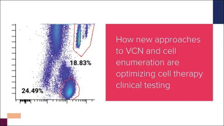
We delve into how clinical testing is evolving to meet demand in this rapidly changing landscape in our Cell Therapy Trends Report, and examine a few key areas where cell therapy testing programs are changing. One of these is cell enumeration and vector copy number (VCN) determination, which provide insights on how a given therapeutic construct is behaving in the body.
Below, we explore how techniques for cell enumeration and VCN determination are being adapted to assess greater numbers of increasingly complex and diversified therapy candidates, and discuss what the future holds for this key area of clinical testing.
Cell enumeration and VCN determination methods assess cell number (via flow cytometric detection of surface proteins) and the number of constructs transduced into a cell (via PCR-based quantification of DNA), respectively. Together, the two measure cell therapy expansion and determine the abundance and persistence of therapeutic cells — crucial to ascertain how various cell types are responding to a therapy.
As cell therapies and sample types diversify, clinical testing for cell enumeration and VCN determination must assess a broader array of targets, requiring more specialized means of analysis. However, developers must also keep speed in mind, and balance specialization with efficiency to ensure that therapies are evaluated in a timely way.
So, what does the future hold for this realm of clinical testing?
Cell enumeration and VCN determination techniques take complementary measurements from different angles, using different methods and analytes to evaluate cell therapies.
By utilizing both approaches in early-phase trials, cell therapy developers can gain high confidence in how they are interpreting their data, thereby producing a more reliable and robust picture of therapy expansion, and also lending confidence to each individual assay.
Alignment can also streamline testing. Cell enumeration via flow cytometry, despite providing additional phenotyping information on expanded cells, can be difficult to scale up, and so is often limited in later-stage use due to the challenges of collecting and transporting samples off-site. But, if aligned with cell enumeration in early-phase testing, VCN can act as a proxy measure of cell number during later-stage testing, simplifying logistics.
As more diverse cell therapies reach clinical stages, cell enumeration and VCN determination methods need increasing customization — to track proprietary constructs as patients become more likely to receive multiple therapies, for instance. Additionally, new therapeutic cells such as CAR-macrophages may require novel antigen targets to be monitored in various tissues other than circulating blood, requiring new protocols for collection and analysis.
Although customized assays are valuable, they are time- and resource-consuming to develop and validate. Modularization can minimize the timeline impact of assay customization. Testing programs for cell enumeration and VCN determination can be made quicker and easier to deploy by identifying generic analytes; and by utilizing modular data analytics to streamline readouts, and accelerate data processing.
With the accelerating pace and increasing diversity of cell therapy development, clinical testing must become more responsive and adaptive, leveraging different approaches to deliver readouts that provide meaningful insights into cell therapy action and host response.
We dig deeper into emerging testing approaches for cell enumeration and VCN determination, and explore the future of cell therapy clinical testing more generally, in our Cell Therapy Trends Report. Download the report now, or contact our team to speak to an expert about your cell therapy needs.
About the author:

Laïla-Aïcha Hanafi is the Director of Scientific Laboratory Operations in flow cytometry at CellCarta. She supplemented her PhD in immuno-oncology with post-doctoral studies in translational biomarkers for cell therapies at the Fred Hutchinson Cancer Center. Laïla has combined scientific knowledge and operational efficiency to address biomarker needs in clinical trial and prioritizing high quality data to move therapies to the next stage of clinical deployment.
July 16, 2024
Cell therapy targets, constructs, and agents are expanding and diversifying — fast. Clinical testing must now comprehensively characterize an ever-broader number and array of therapies, while continuing to prioritize urgency and efficiency in their development and approval.
We delve into how clinical testing is evolving to meet demand in this rapidly changing landscape in our Cell Therapy Trends Report, and examine a few key areas where cell therapy testing programs are changing. One of these is cell enumeration and vector copy number (VCN) determination, which provide insights on how a given therapeutic construct is behaving in the body.
Below, we explore how techniques for cell enumeration and VCN determination are being adapted to assess greater numbers of increasingly complex and diversified therapy candidates, and discuss what the future holds for this key area of clinical testing.
Cell enumeration and VCN determination methods assess cell number (via flow cytometric detection of surface proteins) and the number of constructs transduced into a cell (via PCR-based quantification of DNA), respectively. Together, the two measure cell therapy expansion and determine the abundance and persistence of therapeutic cells — crucial to ascertain how various cell types are responding to a therapy.
As cell therapies and sample types diversify, clinical testing for cell enumeration and VCN determination must assess a broader array of targets, requiring more specialized means of analysis. However, developers must also keep speed in mind, and balance specialization with efficiency to ensure that therapies are evaluated in a timely way.
So, what does the future hold for this realm of clinical testing?
Cell enumeration and VCN determination techniques take complementary measurements from different angles, using different methods and analytes to evaluate cell therapies.
By utilizing both approaches in early-phase trials, cell therapy developers can gain high confidence in how they are interpreting their data, thereby producing a more reliable and robust picture of therapy expansion, and also lending confidence to each individual assay.
Alignment can also streamline testing. Cell enumeration via flow cytometry, despite providing additional phenotyping information on expanded cells, can be difficult to scale up, and so is often limited in later-stage use due to the challenges of collecting and transporting samples off-site. But, if aligned with cell enumeration in early-phase testing, VCN can act as a proxy measure of cell number during later-stage testing, simplifying logistics.
As more diverse cell therapies reach clinical stages, cell enumeration and VCN determination methods need increasing customization — to track proprietary constructs as patients become more likely to receive multiple therapies, for instance. Additionally, new therapeutic cells such as CAR-macrophages may require novel antigen targets to be monitored in various tissues other than circulating blood, requiring new protocols for collection and analysis.
Although customized assays are valuable, they are time- and resource-consuming to develop and validate. Modularization can minimize the timeline impact of assay customization. Testing programs for cell enumeration and VCN determination can be made quicker and easier to deploy by identifying generic analytes; and by utilizing modular data analytics to streamline readouts, and accelerate data processing.
With the accelerating pace and increasing diversity of cell therapy development, clinical testing must become more responsive and adaptive, leveraging different approaches to deliver readouts that provide meaningful insights into cell therapy action and host response.
We dig deeper into emerging testing approaches for cell enumeration and VCN determination, and explore the future of cell therapy clinical testing more generally, in our Cell Therapy Trends Report. Download the report now, or contact our team to speak to an expert about your cell therapy needs.
About the author:
Laïla-Aïcha Hanafi is the Director of Scientific Laboratory Operations in flow cytometry at CellCarta. She supplemented her PhD in immuno-oncology with post-doctoral studies in translational biomarkers for cell therapies at the Fred Hutchinson Cancer Center. Laïla has combined scientific knowledge and operational efficiency to address biomarker needs in clinical trial and prioritizing high quality data to move therapies to the next stage of clinical deployment.
October 19, 2022

CellCarta is your one partner for your adoptive cell therapy program by offering a complete suite of services with complementary platforms:
Download our case study on digital PCR.
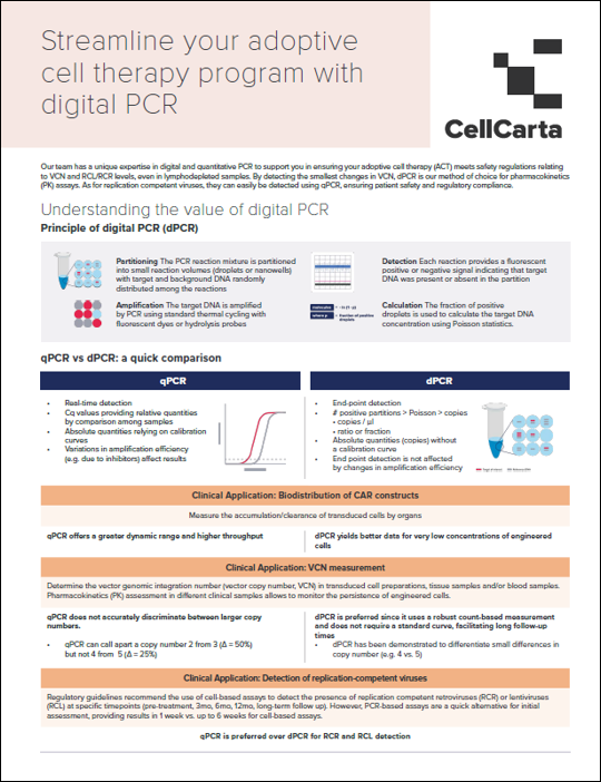
Our team has unique expertise in digital PCR to support you in ensuring your adoptive cell therapy meets safety regulations relating to vector copy number (VCN).
Download the case study for an overview on dPCR:
As a global Contract Research Organization Laboratory (CRO) to the biopharmaceutical industry, CellCarta provides access to a broad offering of biomarker platforms and services. We partner with you to address the most complex laboratory testing needs, delivering customized biomarker testing solutions to further the limitless potential of precision medicine.
October 19, 2022
CellCarta is your one partner for your adoptive cell therapy program by offering a complete suite of services with complementary platforms:
Download our case study on digital PCR.
Our team has unique expertise in digital PCR to support you in ensuring your adoptive cell therapy meets safety regulations relating to vector copy number (VCN).
Download the case study for an overview on dPCR:
As a global Contract Research Organization Laboratory (CRO) to the biopharmaceutical industry, CellCarta provides access to a broad offering of biomarker platforms and services. We partner with you to address the most complex laboratory testing needs, delivering customized biomarker testing solutions to further the limitless potential of precision medicine.