Understanding the FDA’s Guidance for ADC Development

October 24, 2025

Antibody-drug conjugates (ADCs) have emerged as one of the most promising modalities in oncology treatment, and with more than 15 ADCs approved by the FDA (as of May 2025), they are now firmly established as a proven therapeutic class. To support the growing number of ADCs in development, the FDA issued its first dedicated guidance on clinical pharmacology considerations for ADCs in March 2024, bringing together decades of experience to provide developers with a clearer framework for evaluating these complex therapies throughout clinical development.1,2

So what does this guidance mean for ADC development? And what do developers need to do to meet these expectations

The FDA guidance: understanding the ADC as a sum of its parts1

The new FDA guidance makes it clear that ADCs must be evaluated as multi-component products, as the antibody, the payload, the linker, and any relevant metabolites all contribute to overall safety and efficacy. Evaluations across a clinical pharmacology program must therefore account for the contribution of each component, not just the ADC as a whole. As a result, sponsors are expected to measure each element with validated assays throughout ADC development and provide justification if any are excluded.

The guidance sets expectations across all the core areas of clinical pharmacology: bioanalytical approach, dose– and exposure–response analysis, intrinsic factors, QTc assessment, immunogenicity, and drug–drug interactions. Within this framework, the evaluation of intrinsic factors is a critical consideration, as it captures how patient characteristics, including genetic variation, may influence the behavior of ADC components.

Intrinsic factors and pharmacogenomic considerations

A notable inclusion in the intrinsic factors assessment is the expectation to evaluate pharmacogenomics. The FDA highlights that patient genetics can influence ADC exposure and response—for example, functional variants of enzymes and transporters such as CYP2D6 or BCRP can alter clearance of the unconjugated payload, while Fc-gamma receptor (FcγR) variants may affect antibody-mediated activity. Depending on an ADC’s mechanism, and absorption, distribution, metabolism, and excretion (ADME) profile, a pharmacogenetic evaluation may be recommended to capture these influences.

One area where pharmacogenomics may be particularly relevant is linker stability. ADC payload release relies on cleavage of the chemical linker, a process that can be mediated by enzymes, reduction, or pH-dependent hydrolysis. Reflecting this, the FDA calls out linker-derived analytes in specific assessments, such as QTc risk evaluation, where sponsors are expected to analyze the impact of the unconjugated payload, metabolites, and the linker itself. In practice, this means developers may need to incorporate pharmacogenomic data into studies that examine payload release and exposure, ensuring they capture the impact of patient variability on this critical step of ADC function.

What does the pharmacogenomics guidance mean for ADC development?

For ADC developers, the guidance raises the bar on clinical pharmacology planning. Programs should now:

  • Expand bioanalytical coverage to support pharmacogenomic analyses, with assays that assess, not just the intact ADC, but also the antibody, linker, payload, and relevant metabolites, with clear justification if any are excluded
  • Build pharmacogenomics early into study design, particularly where genetic variants may affect payload clearance or antibody function
  • Account for linker stability in pharmacogenomic assessments, including specialized studies such as QTc evaluations, where regulators expect to see data on both payload and linker analytes

Meeting these expectations will require more detailed planning and earlier integration of component-level assessments into ADC development programs. Teams that prepare for these requirements early can reduce regulatory risk and maintain momentum through development.

Find out more about how CellCarta can support your ADC development programs.

About the author:

author photo

Nathalie Bernard (PhD) is the scientific business director for the Genomic Services unit within CellCarta. Her background is in molecular biology, and she has many years of experience in PCR and sequencing, technologies used to discover or identify DNA and RNA biomarkers of clinical utility. At CellCarta, Nathalie is using her expertise to guide our customers in finding the best solution to their genomic questions.

References

  1. https://www.fda.gov/regulatory-information/search-fda-guidance-documents/clinical-pharmacology-considerations-antibody-drug-conjugates-guidance-industry  
  2. https://www.fda.gov/drugs/guidances-drugs/guidance-recap-podcast-clinical-pharmacology-considerations-antibody-drug-conjugates   

Understanding the FDA’s Guidance for ADC Development

October 24, 2025

Antibody-drug conjugates (ADCs) have emerged as one of the most promising modalities in oncology treatment, and with more than 15 ADCs approved by the FDA (as of May 2025), they are now firmly established as a proven therapeutic class. To support the growing number of ADCs in development, the FDA issued its first dedicated guidance on clinical pharmacology considerations for ADCs in March 2024, bringing together decades of experience to provide developers with a clearer framework for evaluating these complex therapies throughout clinical development.1,2

So what does this guidance mean for ADC development? And what do developers need to do to meet these expectations

The FDA guidance: understanding the ADC as a sum of its parts1

The new FDA guidance makes it clear that ADCs must be evaluated as multi-component products, as the antibody, the payload, the linker, and any relevant metabolites all contribute to overall safety and efficacy. Evaluations across a clinical pharmacology program must therefore account for the contribution of each component, not just the ADC as a whole. As a result, sponsors are expected to measure each element with validated assays throughout ADC development and provide justification if any are excluded.

The guidance sets expectations across all the core areas of clinical pharmacology: bioanalytical approach, dose– and exposure–response analysis, intrinsic factors, QTc assessment, immunogenicity, and drug–drug interactions. Within this framework, the evaluation of intrinsic factors is a critical consideration, as it captures how patient characteristics, including genetic variation, may influence the behavior of ADC components.

Intrinsic factors and pharmacogenomic considerations

A notable inclusion in the intrinsic factors assessment is the expectation to evaluate pharmacogenomics. The FDA highlights that patient genetics can influence ADC exposure and response—for example, functional variants of enzymes and transporters such as CYP2D6 or BCRP can alter clearance of the unconjugated payload, while Fc-gamma receptor (FcγR) variants may affect antibody-mediated activity. Depending on an ADC’s mechanism, and absorption, distribution, metabolism, and excretion (ADME) profile, a pharmacogenetic evaluation may be recommended to capture these influences.

One area where pharmacogenomics may be particularly relevant is linker stability. ADC payload release relies on cleavage of the chemical linker, a process that can be mediated by enzymes, reduction, or pH-dependent hydrolysis. Reflecting this, the FDA calls out linker-derived analytes in specific assessments, such as QTc risk evaluation, where sponsors are expected to analyze the impact of the unconjugated payload, metabolites, and the linker itself. In practice, this means developers may need to incorporate pharmacogenomic data into studies that examine payload release and exposure, ensuring they capture the impact of patient variability on this critical step of ADC function.

What does the pharmacogenomics guidance mean for ADC development?

For ADC developers, the guidance raises the bar on clinical pharmacology planning. Programs should now:

  • Expand bioanalytical coverage to support pharmacogenomic analyses, with assays that assess, not just the intact ADC, but also the antibody, linker, payload, and relevant metabolites, with clear justification if any are excluded
  • Build pharmacogenomics early into study design, particularly where genetic variants may affect payload clearance or antibody function
  • Account for linker stability in pharmacogenomic assessments, including specialized studies such as QTc evaluations, where regulators expect to see data on both payload and linker analytes

Meeting these expectations will require more detailed planning and earlier integration of component-level assessments into ADC development programs. Teams that prepare for these requirements early can reduce regulatory risk and maintain momentum through development.

Find out more about how CellCarta can support your ADC development programs.

About the author:

author photo

Nathalie Bernard (PhD) is the scientific business director for the Genomic Services unit within CellCarta. Her background is in molecular biology, and she has many years of experience in PCR and sequencing, technologies used to discover or identify DNA and RNA biomarkers of clinical utility. At CellCarta, Nathalie is using her expertise to guide our customers in finding the best solution to their genomic questions.
References

  1. https://www.fda.gov/regulatory-information/search-fda-guidance-documents/clinical-pharmacology-considerations-antibody-drug-conjugates-guidance-industry  
  2. https://www.fda.gov/drugs/guidances-drugs/guidance-recap-podcast-clinical-pharmacology-considerations-antibody-drug-conjugates   

Tregs in Focus: How a Nobel-Winning Finding is Shaping Clinical Research

October 22, 2025

The 2025 Nobel Prize in Physiology or Medicine recognized discoveries that transformed our understanding of how the immune system maintains balance. Mary Brunkow, Frederick Ramsdell, and Shimon Sakaguchi were awarded for uncovering the mechanisms of peripheral immune tolerance, a process that prevents our immune system from unduly attacking the body’s own tissues.¹  

Through decades of research, they revealed that immune tolerance is not just established during T-cell development in the thymus but is actively maintained throughout life by a specialized population of FOXP3 regulatory T cells (Tregs). Their research demonstrated that Treg cells act as the immune system’s ‘braking mechanism’, suppressing inappropriate or excessive activation that could harm the body.¹  

Their discoveries bridged a gap that had long puzzled immunologists, explaining how the immune system is dynamically self-regulating throughout life to distinguish between harmful invaders and healthy cells. That knowledge paved the way to today’s wave of Treg-focused clinical trials, where researchers are investigating how tipping the immune balance one way or the other can be achieved through precise manipulation of Treg cells.   

What is the current focus of Treg clinical trials? 

Building on these foundational discoveries, current clinical research is focused on how modulating Treg activity can improve outcomes in patients with cancer and autoimmune diseases. 

Oncology 

In oncology, Treg clinical trials are exploring ways to reduce the suppressive activity of Tregs within the tumor microenvironment (TME), where these cells often accumulate and inhibit anti-tumor immunity.2

Key approaches under investigation include: 

  • Checkpoint and cytokine modulation, such as combining PD-(L)1 and VEGF inhibitors, which can alter the TME to reduce immune suppression, including Treg activity2
  • Blocking chemokine receptors, using antibodies that target CCR4 or CCR8 to prevent Tregs from migrating into the TME and deplete them2
  • Targeted depletion, including next-generation antibody–drug conjugates (ADCs) designed to selectively eliminate Tregs through markers such as CD253 

Early-phase studies show that these strategies can enhance cytotoxic T-cell activity and improve responses to checkpoint inhibitors.1,2  Further research is focused on improving selectivity, ensuring that Tregs in the TME can be modulated without disrupting the peripheral populations needed to maintain overall immune balance.2 

Autoimmune diseases 

In autoimmune and inflammatory disorders, clinical strategies take the opposite approach by enhancing or stabilizing Tregs to re-establish immune tolerance.  

Current research includes:  

  • Cytokine-based stimulation, using low-dose or engineered interleukin-2 (IL-2) therapies that selectively expand Tregs while avoiding activation of effector T cells4,5
  • Adoptive or engineered Treg therapies, such as CAR-Tregs and T-cell receptor (TCR)-Tregs, which are being developed to deliver site-specific immune suppression and promote graft tolerance in transplantation4,5 

Early clinical data suggest that these approaches can expand functional Tregs and reduce autoimmune inflammation without broadly suppressing protective immunity. As research progresses, the aim is to improve the scalability, safety, and efficacy of Treg cell therapies to make them more accessible to a broader patient population.5 

How are Tregs assessed in clinical trials?  

Accurate measurement of Tregs is essential for evaluating treatment effects in clinical studies. Assessments are typically performed in both tissue and blood samples, each providing complementary insight into Treg frequency, localization, and function. 

Assessment in tissues  

Tissue analysis provides spatial information about where Tregs are located and how they interact with other immune cells within the tissue microenvironment. Biomarkers such as FOXP3, CD3, and CD4 are commonly used to identify Tregs in histopathology samples. To capture this in high resolution, researchers can use multiplex imaging platforms such as the Lunaphore COMET™ system to detect dozens of markers at single-cell resolution, enabling high-plex visualization of Tregs and their surrounding context. 

Assessment in blood  

Blood-based analysis focuses on circulating Tregs, typically defined by CD25 and CD127 expression, with FOXP3 used as a confirmatory marker. Flow cytometry can be used to quantify Treg frequency and phenotype in whole blood or isolated peripheral blood mononuclear cells (PBMCs). 

What are the challenges of assessing Tregs in clinical trials?  

While these analytical methods are well established, accurately quantifying Tregs in clinical samples can be challenging. Their low frequency and sensitivity to handling, in addition to a reliance on intracellular markers such as FOXP3 mean that even minor differences in sample processing and methodology can impact results.  

Sample stability and processing 

Tregs are highly sensitive to sample handling, which can make them difficult to assess accurately . During PBMC isolation, there can be up to a fivefold decrease in Treg populations, even when samples are processed within 24 hours of blood draw.        

To avoid this, researchers can use CytoChex® blood collection tubes for whole blood collection if Tregs are a key readout, as this approach has been found to better preserve Treg cell frequency and marker expression over time.  

Staining resolution 

Because FOXP3 is an intracellular marker, accurate detection depends on optimized fixation, permeabilization, and gating. Inconsistent preparation may blur signal distinction and complicate gating. 

This can be addressed through optimized staining protocols and refined gating strategies, which can improve the reproducibility and clarity of FOXP3 Treg readouts in whole blood.  

Treg heterogeneity 

Tregs are not a uniform population. Differences in stability, activation state, and function can complicate data interpretation, particularly when bulk assays average out signals across diverse subsets. 

This complexity can be addressed through single-cell transcriptomic and TCR-sequencing approaches, which can identify distinct Treg subpopulations and track how individual clones shift in phenotype or functionality over time. These methods provide a more detailed view of Treg diversity and its relevance to therapeutic response. 

Looking ahead 

The discoveries recognized by the 2025 Nobel Prize underpin an expanding focus on Tregs in clinical immunology and oncology research. As therapeutic programs increasingly incorporate Treg assessment, advances in sample stabilization, staining workflows, and multi-omic profiling can help deliver more consistent, high-resolution data to help researchers characterize these cells and link their activity to clinical outcomes. 

These deeper insights can help pave the way for more precise, effective therapies that could offer new potential to treat cancer and autoimmune diseases. 

Contact us to find out how CellCarta’s immunology and biomarker expertise can support Treg assessment and analysis.  

About the author:

author photo

Céline Vandamme is a Scientific Business Director at CellCarta, specializing in the flow cytometry platform. With a PhD in immunology, and a broad expertise gained through her work at various academic and pharmaceutical institutions, Céline has profuse experience in designing flow cytometry assays to support immune monitoring activities in clinical trials.

References 

  1. https://www.nobelprize.org/prizes/medicine/2025/popular-information/ 
  2. https://pmc.ncbi.nlm.nih.gov/articles/PMC9588644/#:~:text=
    There%20are%20seven%20proposed%20methods,Treg%20cytokine%20secretion%2C%20(6)
      
  3. https://www.nature.com/articles/s12276-023-01080-3  
  4. https://www.frontiersin.org/journals/immunology/articles/10.3389/fimmu.2025.1511671/full  
  5. https://www.delveinsight.com/blog/treg-cell-based-therapies-in-pipeline 

Tregs in Focus: How a Nobel-Winning Finding is Shaping Clinical Research

October 22, 2025

The 2025 Nobel Prize in Physiology or Medicine recognized discoveries that transformed our understanding of how the immune system maintains balance. Mary Brunkow, Frederick Ramsdell, and Shimon Sakaguchi were awarded for uncovering the mechanisms of peripheral immune tolerance, a process that prevents our immune system from unduly attacking the body’s own tissues.¹  

Through decades of research, they revealed that immune tolerance is not just established during T-cell development in the thymus but is actively maintained throughout life by a specialized population of FOXP3 regulatory T cells (Tregs). Their research demonstrated that Treg cells act as the immune system’s ‘braking mechanism’, suppressing inappropriate or excessive activation that could harm the body.¹  

Their discoveries bridged a gap that had long puzzled immunologists, explaining how the immune system is dynamically self-regulating throughout life to distinguish between harmful invaders and healthy cells. That knowledge paved the way to today’s wave of Treg-focused clinical trials, where researchers are investigating how tipping the immune balance one way or the other can be achieved through precise manipulation of Treg cells.   

What is the current focus of Treg clinical trials? 

Building on these foundational discoveries, current clinical research is focused on how modulating Treg activity can improve outcomes in patients with cancer and autoimmune diseases. 

Oncology 

In oncology, Treg clinical trials are exploring ways to reduce the suppressive activity of Tregs within the tumor microenvironment (TME), where these cells often accumulate and inhibit anti-tumor immunity.2

Key approaches under investigation include: 

  • Checkpoint and cytokine modulation, such as combining PD-(L)1 and VEGF inhibitors, which can alter the TME to reduce immune suppression, including Treg activity2
  • Blocking chemokine receptors, using antibodies that target CCR4 or CCR8 to prevent Tregs from migrating into the TME and deplete them2
  • Targeted depletion, including next-generation antibody–drug conjugates (ADCs) designed to selectively eliminate Tregs through markers such as CD253 

Early-phase studies show that these strategies can enhance cytotoxic T-cell activity and improve responses to checkpoint inhibitors.1,2  Further research is focused on improving selectivity, ensuring that Tregs in the TME can be modulated without disrupting the peripheral populations needed to maintain overall immune balance.2 

Autoimmune diseases 

In autoimmune and inflammatory disorders, clinical strategies take the opposite approach by enhancing or stabilizing Tregs to re-establish immune tolerance.  

Current research includes:  

  • Cytokine-based stimulation, using low-dose or engineered interleukin-2 (IL-2) therapies that selectively expand Tregs while avoiding activation of effector T cells4,5
  • Adoptive or engineered Treg therapies, such as CAR-Tregs and T-cell receptor (TCR)-Tregs, which are being developed to deliver site-specific immune suppression and promote graft tolerance in transplantation4,5 

Early clinical data suggest that these approaches can expand functional Tregs and reduce autoimmune inflammation without broadly suppressing protective immunity. As research progresses, the aim is to improve the scalability, safety, and efficacy of Treg cell therapies to make them more accessible to a broader patient population.5 

How are Tregs assessed in clinical trials?  

Accurate measurement of Tregs is essential for evaluating treatment effects in clinical studies. Assessments are typically performed in both tissue and blood samples, each providing complementary insight into Treg frequency, localization, and function. 

Assessment in tissues  

Tissue analysis provides spatial information about where Tregs are located and how they interact with other immune cells within the tissue microenvironment. Biomarkers such as FOXP3, CD3, and CD4 are commonly used to identify Tregs in histopathology samples. To capture this in high resolution, researchers can use multiplex imaging platforms such as the Lunaphore COMET™ system to detect dozens of markers at single-cell resolution, enabling high-plex visualization of Tregs and their surrounding context. 

Assessment in blood  

Blood-based analysis focuses on circulating Tregs, typically defined by CD25 and CD127 expression, with FOXP3 used as a confirmatory marker. Flow cytometry can be used to quantify Treg frequency and phenotype in whole blood or isolated peripheral blood mononuclear cells (PBMCs). 

What are the challenges of assessing Tregs in clinical trials?  

While these analytical methods are well established, accurately quantifying Tregs in clinical samples can be challenging. Their low frequency and sensitivity to handling, in addition to a reliance on intracellular markers such as FOXP3 mean that even minor differences in sample processing and methodology can impact results.  

Sample stability and processing 

Tregs are highly sensitive to sample handling, which can make them difficult to assess accurately . During PBMC isolation, there can be up to a fivefold decrease in Treg populations, even when samples are processed within 24 hours of blood draw.        

To avoid this, researchers can use CytoChex® blood collection tubes for whole blood collection if Tregs are a key readout, as this approach has been found to better preserve Treg cell frequency and marker expression over time.  

Staining resolution 

Because FOXP3 is an intracellular marker, accurate detection depends on optimized fixation, permeabilization, and gating. Inconsistent preparation may blur signal distinction and complicate gating. 

This can be addressed through optimized staining protocols and refined gating strategies, which can improve the reproducibility and clarity of FOXP3 Treg readouts in whole blood.  

Treg heterogeneity 

Tregs are not a uniform population. Differences in stability, activation state, and function can complicate data interpretation, particularly when bulk assays average out signals across diverse subsets. 

This complexity can be addressed through single-cell transcriptomic and TCR-sequencing approaches, which can identify distinct Treg subpopulations and track how individual clones shift in phenotype or functionality over time. These methods provide a more detailed view of Treg diversity and its relevance to therapeutic response. 

Looking ahead 

The discoveries recognized by the 2025 Nobel Prize underpin an expanding focus on Tregs in clinical immunology and oncology research. As therapeutic programs increasingly incorporate Treg assessment, advances in sample stabilization, staining workflows, and multi-omic profiling can help deliver more consistent, high-resolution data to help researchers characterize these cells and link their activity to clinical outcomes. 

These deeper insights can help pave the way for more precise, effective therapies that could offer new potential to treat cancer and autoimmune diseases. 

Contact us to find out how CellCarta’s immunology and biomarker expertise can support Treg assessment and analysis.  

About the author:

author photo

Céline Vandamme is a Scientific Business Director at CellCarta, specializing in the flow cytometry platform. With a PhD in immunology, and a broad expertise gained through her work at various academic and pharmaceutical institutions, Céline has profuse experience in designing flow cytometry assays to support immune monitoring activities in clinical trials.

References 

  1. https://www.nobelprize.org/prizes/medicine/2025/popular-information/ 
  2. https://pmc.ncbi.nlm.nih.gov/articles/PMC9588644/#:~:text=
    There%20are%20seven%20proposed%20methods,Treg%20cytokine%20secretion%2C%20(6)
      
  3. https://www.nature.com/articles/s12276-023-01080-3  
  4. https://www.frontiersin.org/journals/immunology/articles/10.3389/fimmu.2025.1511671/full  
  5. https://www.delveinsight.com/blog/treg-cell-based-therapies-in-pipeline 

B-Cell Populations in Focus: A New Flow Cytometry Assay for Increased Sensitivity in Autoimmune Research

September 19, 2025

B cells are central to both protective immunity and autoimmune pathology. With the recent shift of cell therapies from oncology indications to autoimmune diseases, interest has been growing towards this immune cell population.1,2

Flow cytometry remains the gold standard for monitoring B cells in both research and clinical settings. However, lack of convention for classification as well as reliable and stable markers, especially following thawing of samples, can make it difficult to capture rare B-cell populations relevant to autoimmune drug development.

For translational research teams, this creates an urgent need for tools that deliver the sensitivity and consistency required to fully understand B-cell dynamics and drive the development of new B-cell therapies.

The Challenge of Defining B-cell Subsets

B cells are far from uniform. Transitional, naïve, memory, double-negative, and plasma cell subsets each play distinct roles in health and disease, but their phenotypic profiles often overlap. Inconsistent marker usage and variability in gating strategies have made it difficult to classify B-cell subsets consistently, which can complicate the comparison of results across studies.3

For drug developers, the lack of standardization creates real-world hurdles. Autoimmune therapies that aim to induce a B-cell reset (eliminating autoreactive populations and allowing the immune system to repopulate with naïve B cells) depend on precise monitoring of which subsets return after the B-cell aplasia phase. Without consistent, high-resolution phenotyping, it is difficult to determine whether a therapy is resetting B-cell populations in a way that supports durable disease control.

Recent efforts to harmonize approaches are beginning to address these issues, with publications recommending standardized marker sets to improve reproducibility and resolution of B-cell subsets.³ Markers such as CD21, once regarded mainly for their functional role, are now recognized as important phenotypic indicators that help distinguish distinct populations. Incorporating markers like these into flow cytometry panels is essential to provide the clarity and consistency needed for research on B-cell reset in autoimmune diseases.

A Refined Flow Cytometry Assay for B-Cell Subsets

To address these challenges, CellCarta has advanced its flow cytometry capabilities with a refined B-cell assay that improves the sensitivity and consistency of B-cell subset profiling.

1- Updated gating strategy

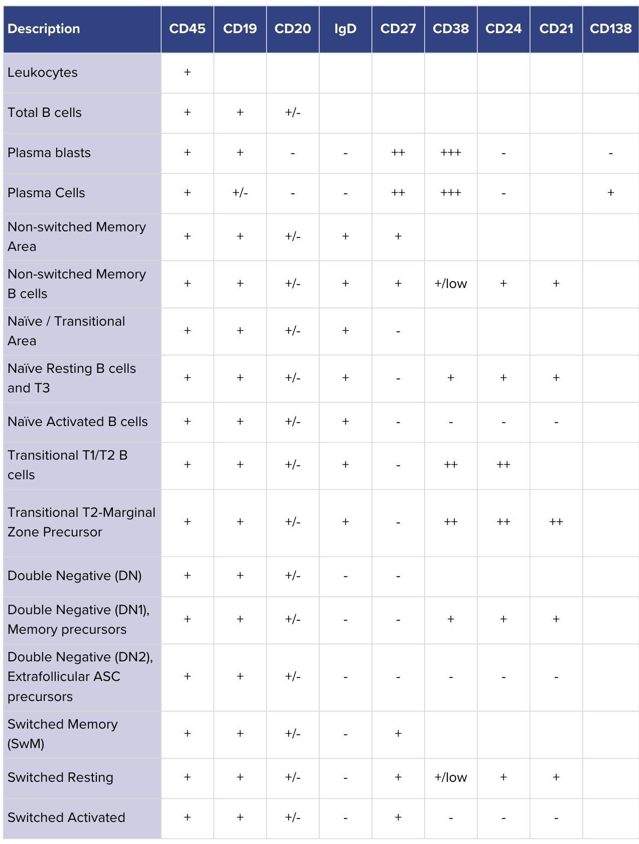
We refined our panel strategy by incorporating markers such as CD21 as phenotypic indicators alongside other established markers. This enables further stratification of B-cell subtypes, providing better insight into both activated and developmentally distinct populations that are increasingly linked to autoimmune disease.

Section image

Table 1: B-cell subsets identifiable with CellCarta’s refined flow cytometry panel. Rows show major populations, while columns list the markers used to classify them. CD21 provides added resolution by distinguishing activated and developmentally distinct subsets relevant to autoimmune disease.

2- Improved bulk lysis method

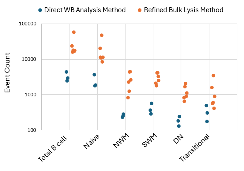
We’ve developed a refined bulk lysis method using the B-cell panel. In comparison to the direct whole blood (WB) analysis (100ul of blood), the improved bulk lysis method resulted in a 10-fold increase in the cell input compared to the traditional analysis using WB for flow cytometry assay (Figure 1). As more cells are acquired, rare populations can now be assessed with better precision. The whole method was evaluated at both 4°C and room temperature (RT), providing flexibility for the sample management and transportation.

Figure 1: Event counts of total B cells and subsets acquired from healthy donors using direct whole blood analysis method (blue) and refined bulk lysis method (orange) WB – Whole Blood; NWM – Non-switched memory; SWM – Switched memory, DN – Double negative

Global Scalability With the Lyric Platform

To offer global site-to-site consistency, we switched our B-cell assay from the Fortessa to the Lyric platform, leveraging the built-in capabilities of this platform to achieve high inter-instrument/inter-site standardization. Unified workflows and directly comparable data make it easier to scale studies internationally, reduce variability, and build confidence in results across multicenter trials.

Supporting the Next Generation of B-Cell Therapies

With refinements to its gating strategy, bulk lysis method, and global platform standardization, CellCarta’s B-cell flow cytometry assay provides the resolution, sensitivity, and reproducibility needed to study B-cell dynamics in detail. In the context of the development of B-cell reset therapies, these advancements enable the precise monitoring and consistency needed to better define which B-cell populations are depleted, which repopulate, and how these shifts influence autoimmune disease outcomes.

In addition to our B-cell panel, CellCarta can support researchers with both off-the-shelf panels for rapid deployment and custom panel development tailored to specific scientific questions. With this flexibility, pharma teams can choose the right approach for their needs, supporting clearer decision-making in the development of new B–cell–targeted therapies.

Meet our expert:

author photo

Alex Guo, PhD, is a Principal Scientist at CellCarta, specializing in immune monitoring and proteomics. With a doctorate in Immunology and Molecular Oncology, he has extensive expertise in developing and validating novel assays to address complex clinical needs. Alex has led immune monitoring analyses across multiple clinical programs, translating high-dimensional data into actionable insights for clients. He combines deep scientific expertise with practical project execution to advance translational and clinical research.

References

  1. Lee, D. S., Rojas, O. L., & Gommerman, J. L. (2021). Nature reviews Drug discovery, 20(3), 179-199.
  2. Harrison C. Nature Biotechnology, vol. 42, 2024, pp. 995–997.
  3. Sanz, I., Wei, C., Jenks, S. A. et al (2019). Frontiers in immunology10, 2458.

B-Cell Populations in Focus: A New Flow Cytometry Assay for Increased Sensitivity in Autoimmune Research

September 19, 2025

B cells are central to both protective immunity and autoimmune pathology. With the recent shift of cell therapies from oncology indications to autoimmune diseases, interest has been growing towards this immune cell population.1,2

Flow cytometry remains the gold standard for monitoring B cells in both research and clinical settings. However, lack of convention for classification as well as reliable and stable markers, especially following thawing of samples, can make it difficult to capture rare B-cell populations relevant to autoimmune drug development.

For translational research teams, this creates an urgent need for tools that deliver the sensitivity and consistency required to fully understand B-cell dynamics and drive the development of new B-cell therapies.

The Challenge of Defining B-cell Subsets

B cells are far from uniform. Transitional, naïve, memory, double-negative, and plasma cell subsets each play distinct roles in health and disease, but their phenotypic profiles often overlap. Inconsistent marker usage and variability in gating strategies have made it difficult to classify B-cell subsets consistently, which can complicate the comparison of results across studies.3

For drug developers, the lack of standardization creates real-world hurdles. Autoimmune therapies that aim to induce a B-cell reset (eliminating autoreactive populations and allowing the immune system to repopulate with naïve B cells) depend on precise monitoring of which subsets return after the B-cell aplasia phase. Without consistent, high-resolution phenotyping, it is difficult to determine whether a therapy is resetting B-cell populations in a way that supports durable disease control.

Recent efforts to harmonize approaches are beginning to address these issues, with publications recommending standardized marker sets to improve reproducibility and resolution of B-cell subsets.³ Markers such as CD21, once regarded mainly for their functional role, are now recognized as important phenotypic indicators that help distinguish distinct populations. Incorporating markers like these into flow cytometry panels is essential to provide the clarity and consistency needed for research on B-cell reset in autoimmune diseases.

A Refined Flow Cytometry Assay for B-Cell Subsets

To address these challenges, CellCarta has advanced its flow cytometry capabilities with a refined B-cell assay that improves the sensitivity and consistency of B-cell subset profiling.

1- Updated gating strategy

We refined our panel strategy by incorporating markers such as CD21 as phenotypic indicators alongside other established markers. This enables further stratification of B-cell subtypes, providing better insight into both activated and developmentally distinct populations that are increasingly linked to autoimmune disease.

Section image

Table 1: B-cell subsets identifiable with CellCarta’s refined flow cytometry panel. Rows show major populations, while columns list the markers used to classify them. CD21 provides added resolution by distinguishing activated and developmentally distinct subsets relevant to autoimmune disease.

2- Improved bulk lysis method

We’ve developed a refined bulk lysis method using the B-cell panel. In comparison to the direct whole blood (WB) analysis (100ul of blood), the improved bulk lysis method resulted in a 10-fold increase in the cell input compared to the traditional analysis using WB for flow cytometry assay (Figure 1). As more cells are acquired, rare populations can now be assessed with better precision. The whole method was evaluated at both 4°C and room temperature (RT), providing flexibility for the sample management and transportation.

Figure 1: Event counts of total B cells and subsets acquired from healthy donors using direct whole blood analysis method (blue) and refined bulk lysis method (orange) WB – Whole Blood; NWM – Non-switched memory; SWM – Switched memory, DN – Double negative

Global Scalability With the Lyric Platform

To offer global site-to-site consistency, we switched our B-cell assay from the Fortessa to the Lyric platform, leveraging the built-in capabilities of this platform to achieve high inter-instrument/inter-site standardization. Unified workflows and directly comparable data make it easier to scale studies internationally, reduce variability, and build confidence in results across multicenter trials.

Supporting the Next Generation of B-Cell Therapies

With refinements to its gating strategy, bulk lysis method, and global platform standardization, CellCarta’s B-cell flow cytometry assay provides the resolution, sensitivity, and reproducibility needed to study B-cell dynamics in detail. In the context of the development of B-cell reset therapies, these advancements enable the precise monitoring and consistency needed to better define which B-cell populations are depleted, which repopulate, and how these shifts influence autoimmune disease outcomes.

In addition to our B-cell panel, CellCarta can support researchers with both off-the-shelf panels for rapid deployment and custom panel development tailored to specific scientific questions. With this flexibility, pharma teams can choose the right approach for their needs, supporting clearer decision-making in the development of new B–cell–targeted therapies.

Meet our expert:

author photo

Alex Guo, PhD, is a Principal Scientist at CellCarta, specializing in immune monitoring and proteomics. With a doctorate in Immunology and Molecular Oncology, he has extensive expertise in developing and validating novel assays to address complex clinical needs. Alex has led immune monitoring analyses across multiple clinical programs, translating high-dimensional data into actionable insights for clients. He combines deep scientific expertise with practical project execution to advance translational and clinical research.

References

  1. Lee, D. S., Rojas, O. L., & Gommerman, J. L. (2021). Nature reviews Drug discovery, 20(3), 179-199.
  2. Harrison C. Nature Biotechnology, vol. 42, 2024, pp. 995–997.
  3. Sanz, I., Wei, C., Jenks, S. A. et al (2019). Frontiers in immunology10, 2458.

Preclinical Genomic Insights for Therapeutic Development

August 18, 2025

The risk of poor target selection in preclinical development

In early-stage drug development, there’s often a rush to move promising candidates forward. Tight timelines and competitive pressures can lead to companies having an over-reliance on ‘quantity over quality’ in research and development,hoping to maximize their chances of success by bringing more products into the pipeline.

While this may offer short-term momentum, advancing drug candidates without establishing a strong link between target and disease at the preclinical stages can lead to costly, later-stage setbacks. When targets are poorly defined, programs risk progressing on shaky foundations that can compromise other aspects of the pipeline, from study design and patient selection to commercial potential.

Understanding what makes a target worth pursuing, and how to evaluate it effectively, is key to laying the groundwork for clinical success.

Improving trial outcomes starts with the right target

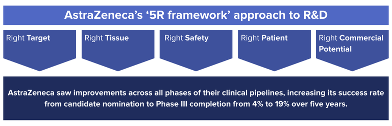
Developing a strong biological foundation for your target is a key driver in the downstream success of a therapeutic candidate. One example of this is AstraZeneca’s ‘5R framework’ approach to R&D, in which they aimed to improve their R&D productivity by focusing on 5 technical determinants: the right target, right tissue, right safety, right patient, and right commercial potential.1

The ‘right target’ principle emphasized selecting targets with a strong link to disease, with a focus on developing a deeper initial biological understanding through fewer, more informative screens—using technologies such as high-content imaging, high-throughput electrophysiology, and advances in genomics technologies such as transcriptomics.

The result? AstraZeneca saw improvements across all phases of their clinical pipelines, increasing its success rate from candidate nomination to Phase III completion from 4% to 19% over five years.1 By focusing only on the most promising candidates and providing robust study data to inform late-stage clinical trials, not only did they improve candidate success rate, they also saved research costs and accelerated all phases of their clinical trials.

Section image

This improvement is a clear demonstration of the impact that strong preclinical research can have on downstream outcomes. A greater focus on target quality in early development enables teams to prioritize candidates with stronger potential for therapeutic impact, increasing the likelihood of success.

The key role of genomics in better target selection

Genomics is a key tool in enabling better target selection, allowing researchers to understand the biological relevance of a target at a molecular level. The growth of techniques in next-generation sequencing (NGS ), RNA sequencing, whole exome sequencing (WES), single-cell sequencing, T-cell/B-cell receptor (TCR/BCR) profiling, and associated analytical methods refining mutation profiling allows teams to uncover associations between genes, pathways, and disease, and assess whether modulating a particular target will likely yield clinical benefit.

In preclinical research, these insights are especially valuable when deciding which candidates to advance, enabling:

  •  Identification of genetically validated targets with known links to human disease
  • Clarification of the mechanism of action (MOA) by showing how a compound influences gene expression or pathway activation
  • Detection of early biomarkers that provide later value in patient stratification or response monitoring
  • Identification of potential off-target effects or resistance mechanisms that could impact long-term viability

Get in touch to find out how we can support your preclinical development.

The impact of early genomic insight on downstream success

The value of genomic profiling doesn’t end in early development. When applied strategically, molecular insights generated in preclinical research can guide key decisions throughout the pipeline.

Early identification of response biomarkers can support more focused trial designs, while understanding resistance pathways can shape monitoring strategies and combination therapy planning. Early-stage genomics work can also lay the foundation for companion diagnostics, enabling the targeted therapy to launch with a clearly defined patient population.

Advances in NGS have led to further investment in genomics to help identify novel, genetically validated targets in early development.1 As such, NGS is now more accessible in early development, allowing teams to build a stronger understanding of target biology and downstream biomarker opportunities sooner in the process.

Case study: Osimertinib and the value of a well-characterized target

The development of Osimertinib, a treatment for non-small cell lung cancer (NSCLC), illustrates how genomic insight can strengthen target confidence and accelerate clinical development.1 In NSCLC, the EGFR T790M mutation had been identified as a common resistance mechanism in patients progressing on first-generation EGFR inhibitors.

AstraZeneca recognized the clinical relevance of this mutation, designing Osimertinib to address this specific, established resistance mechanism. Because the EGFR T790M target was well-characterized, the AstraZeneca team were able to design a more tailored clinical program and use a mutation-specific companion diagnostic for more precise patient selection from the outset.

Combining this with the other elements of the 5R framework resulted in Osimertinib having one of the fastest recorded clinical development programs, going from initial human dosing to launch in just over 2.5 years. The program’s success is a clear example of how well-validated genomic targets can inform trial design, enable targeted patient selection, and accelerate path to market.

Support early development decisions with the right tools and expertise

Translating early genomic insight into clinical success requires the right assays and dedicated expertise. CellCarta offers a wide range of tools suited to preclinical and translational research, including RNAseq, whole genome and whole exome sequencing, single-cell analysis, and Olink proteomics. For companies that require a tailored approach, we offer custom assay development, alongside several validated panels designed around well-established genes, which can streamline trial efficiency. Our validated assay offerings include:

  • oncoReveal® CDx: An FDA-approved targeted NGS panel covering 22 key genes across numerous tumor types
  • TSO500: A comprehensive pan-cancer panel covering 523 genes for DNA variants and 55 for RNA variants
  • Aspyre® lung: Ultra-sensitive, high-fidelity multiplex PCR/reverse-transcriptase (RT)-PCR, covering 11 genes and including a fusion panel for the detection of established NSCLC biomarkers in both DNA and RNA.

By combining these advanced validated platforms with custom analysis pipelines, we can help sponsors narrow down the most promising biomarkers and transition seamlessly into focused platforms, such as qPCR, dPCR, or NGS, at the later clinical trial stages.

Our capabilities can support both small and large pharma companies with early-phase development, right through to regulatory submission. Combined with our global infrastructure, expertise in handling challenging samples, and access to all major genomic analysis platforms, CellCarta ensures high-quality data from early discovery through clinical execution, aiding success at all stages of the pipeline.

Want to learn more about how our services can support your preclinical research? Chat with one of our experts!

References

  1. https://www.nature.com/articles/nrd.2017.244

Preclinical Genomic Insights for Therapeutic Development

August 18, 2025

The risk of poor target selection in preclinical development

In early-stage drug development, there’s often a rush to move promising candidates forward. Tight timelines and competitive pressures can lead to companies having an over-reliance on ‘quantity over quality’ in research and development,hoping to maximize their chances of success by bringing more products into the pipeline.

While this may offer short-term momentum, advancing drug candidates without establishing a strong link between target and disease at the preclinical stages can lead to costly, later-stage setbacks. When targets are poorly defined, programs risk progressing on shaky foundations that can compromise other aspects of the pipeline, from study design and patient selection to commercial potential.

Understanding what makes a target worth pursuing, and how to evaluate it effectively, is key to laying the groundwork for clinical success.

Improving trial outcomes starts with the right target

Developing a strong biological foundation for your target is a key driver in the downstream success of a therapeutic candidate. One example of this is AstraZeneca’s ‘5R framework’ approach to R&D, in which they aimed to improve their R&D productivity by focusing on 5 technical determinants: the right target, right tissue, right safety, right patient, and right commercial potential.1

The ‘right target’ principle emphasized selecting targets with a strong link to disease, with a focus on developing a deeper initial biological understanding through fewer, more informative screens—using technologies such as high-content imaging, high-throughput electrophysiology, and advances in genomics technologies such as transcriptomics.

The result? AstraZeneca saw improvements across all phases of their clinical pipelines, increasing its success rate from candidate nomination to Phase III completion from 4% to 19% over five years.1 By focusing only on the most promising candidates and providing robust study data to inform late-stage clinical trials, not only did they improve candidate success rate, they also saved research costs and accelerated all phases of their clinical trials.

Section image

This improvement is a clear demonstration of the impact that strong preclinical research can have on downstream outcomes. A greater focus on target quality in early development enables teams to prioritize candidates with stronger potential for therapeutic impact, increasing the likelihood of success.

The key role of genomics in better target selection

Genomics is a key tool in enabling better target selection, allowing researchers to understand the biological relevance of a target at a molecular level. The growth of techniques in next-generation sequencing (NGS ), RNA sequencing, whole exome sequencing (WES), single-cell sequencing, T-cell/B-cell receptor (TCR/BCR) profiling, and associated analytical methods refining mutation profiling allows teams to uncover associations between genes, pathways, and disease, and assess whether modulating a particular target will likely yield clinical benefit.

In preclinical research, these insights are especially valuable when deciding which candidates to advance, enabling:

  •  Identification of genetically validated targets with known links to human disease
  • Clarification of the mechanism of action (MOA) by showing how a compound influences gene expression or pathway activation
  • Detection of early biomarkers that provide later value in patient stratification or response monitoring
  • Identification of potential off-target effects or resistance mechanisms that could impact long-term viability

Get in touch to find out how we can support your preclinical development.

The impact of early genomic insight on downstream success

The value of genomic profiling doesn’t end in early development. When applied strategically, molecular insights generated in preclinical research can guide key decisions throughout the pipeline.

Early identification of response biomarkers can support more focused trial designs, while understanding resistance pathways can shape monitoring strategies and combination therapy planning. Early-stage genomics work can also lay the foundation for companion diagnostics, enabling the targeted therapy to launch with a clearly defined patient population.

Advances in NGS have led to further investment in genomics to help identify novel, genetically validated targets in early development.1 As such, NGS is now more accessible in early development, allowing teams to build a stronger understanding of target biology and downstream biomarker opportunities sooner in the process.

Case study: Osimertinib and the value of a well-characterized target

The development of Osimertinib, a treatment for non-small cell lung cancer (NSCLC), illustrates how genomic insight can strengthen target confidence and accelerate clinical development.1 In NSCLC, the EGFR T790M mutation had been identified as a common resistance mechanism in patients progressing on first-generation EGFR inhibitors.

AstraZeneca recognized the clinical relevance of this mutation, designing Osimertinib to address this specific, established resistance mechanism. Because the EGFR T790M target was well-characterized, the AstraZeneca team were able to design a more tailored clinical program and use a mutation-specific companion diagnostic for more precise patient selection from the outset.

Combining this with the other elements of the 5R framework resulted in Osimertinib having one of the fastest recorded clinical development programs, going from initial human dosing to launch in just over 2.5 years. The program’s success is a clear example of how well-validated genomic targets can inform trial design, enable targeted patient selection, and accelerate path to market.

Support early development decisions with the right tools and expertise

Translating early genomic insight into clinical success requires the right assays and dedicated expertise. CellCarta offers a wide range of tools suited to preclinical and translational research, including RNAseq, whole genome and whole exome sequencing, single-cell analysis, and Olink proteomics. For companies that require a tailored approach, we offer custom assay development, alongside several validated panels designed around well-established genes, which can streamline trial efficiency. Our validated assay offerings include:

  • oncoReveal® CDx: An FDA-approved targeted NGS panel covering 22 key genes across numerous tumor types
  • TSO500: A comprehensive pan-cancer panel covering 523 genes for DNA variants and 55 for RNA variants
  • Aspyre® lung: Ultra-sensitive, high-fidelity multiplex PCR/reverse-transcriptase (RT)-PCR, covering 11 genes and including a fusion panel for the detection of established NSCLC biomarkers in both DNA and RNA.

By combining these advanced validated platforms with custom analysis pipelines, we can help sponsors narrow down the most promising biomarkers and transition seamlessly into focused platforms, such as qPCR, dPCR, or NGS, at the later clinical trial stages.

Our capabilities can support both small and large pharma companies with early-phase development, right through to regulatory submission. Combined with our global infrastructure, expertise in handling challenging samples, and access to all major genomic analysis platforms, CellCarta ensures high-quality data from early discovery through clinical execution, aiding success at all stages of the pipeline.

Want to learn more about how our services can support your preclinical research? Chat with one of our experts!

References

  1. https://www.nature.com/articles/nrd.2017.244

The Power of Dual Extraction for Pre-Analytical Samples

August 18, 2025

The value of pre-analytical processes

How samples are initially handled, stored, and utilized can determine how much they are able to contribute to a study later down the line, particularly in longitudinal studies where samples may need to be revisited across multiple timepoints or analyses.

Yet pre-analytical processes, such as sample accessioning and processing, are often regarded simply as technical necessities rather than strategic opportunities to extend sample utility. When done thoughtfully, pre-analytical processes can add significant value, helping teams get more data from less material while preserving data quality.

How early sample management decisions shape long-term success

After a sample is collected, its value depends on the decisions made next. Accessioning, storage, and processing steps each influence how much insight can be generated from a sample, and how consistently, across the course of a study.

During sample accessioning, standardized intake and documentation are key to ensure traceability across sites and timepoints. From there, storage methods must align with both immediate and long-term analysis goals. If these steps aren’t carefully managed, samples may be mislabeled, mishandled, or stored incorrectly, all of which could compromise stability and impact their downstream applications.

If genomic analysis is expected at any stage of the study, it is vital that this is also taken into account at the pre-analytical planning stage. Building it into the sample strategy early can help to maximize the value of each sample and avoid missed opportunities for insight.
DNA/RNA extraction is a pre-analytical step that lays the foundation for reliable downstream analysis by preserving nucleic acid quality and yield, both essential for generating robust molecular data. It is also important to capture molecular quality control (QC) metrics and metadata at this stage to support data integrity and traceability.

However, given the limited availability of patient samples, many teams are hesitant to perform DNA/RNA extraction, and when they do, it’s often done cautiously, extracting DNA and RNA separately out of concern of compromising data quality. In doing so, they may consume more of the sample than necessary, limiting what can be done later in the study.

Optimized and validated dual DNA/RNA extraction, however, can overcome these challenges. Co-extraction techniques can enable high-quality extraction of both analytes from a single sample, reducing material consumption without compromising data quality. More of the original tissue or sample is preserved for future testing, while both DNA and RNA are made available for downstream analysis.

With a validated co-extraction approach in place, and with a CRO partner that has deep sample management expertise, you can gain valuable genomic insights while preserving sample integrity for use throughout the study lifecycle.

Dual Extraction: Get more from your slides

Dual extraction of DNA/RNA generates rich genomic data, immortalizing sample value for use across future analyses.

At CellCarta, we offer dual DNA/RNA tissue extraction supported by pathologists, who are able to calculate the exact number of slides needed to achieve target yields. Using this method, we can reduce the amount of sample required for extraction, so you can be sure that tissue is being used as efficiently as possible and is preserved for future analysis.

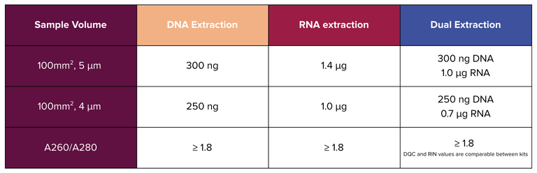
Validation data (Figure 1) shows that our dual extraction method achieves yields comparable to single-analyte extraction kits, demonstrating that high performance can be achieved with less material.

Section image

Figure 1: Yield metrics for DNA and RNA extraction from 4 μm and 5 μm slides, using standard single-analyte kits vs. CellCarta’s dual extraction method (Allprep)

By minimizing sample consumption while maintaining quality, dual extraction enables more from every slide and helps teams carry sample value further through the study.

Speak to one of our experts to find out more about how our dual extraction method can support your next project.

Trusted, end-to-end sample management

Alongside our dual extraction offering, CellCarta’s meticulous sample management and logistics services are designed to protect sample integrity at every step and keep trials running smoothly. We provide:

  • Proven expertise with limited-stability samples; with our expert transport management services, we ensure sample shipping within 24 hours, achieving a 100% on-time study setup
  • Global sample shipping, with capabilities to support trial locations spanning North and South America, Europe, Africa, Asia, and Australia
  • Real-time tracking and reconciliation for full chain-of-custody visibility
  • Dedicated project managers offering personalized oversight from study startup to final analysis

Governed by our robust quality management system, CellCarta helps ensure every sample is moved quickly, reliably, and with full traceability across every stage of your study, so you can get the quality data you need.

Looking to strengthen your pre-analytical strategy? Contact our team to find out how we can support your next study.

The Power of Dual Extraction for Pre-Analytical Samples

August 18, 2025

The value of pre-analytical processes

How samples are initially handled, stored, and utilized can determine how much they are able to contribute to a study later down the line, particularly in longitudinal studies where samples may need to be revisited across multiple timepoints or analyses.

Yet pre-analytical processes, such as sample accessioning and processing, are often regarded simply as technical necessities rather than strategic opportunities to extend sample utility. When done thoughtfully, pre-analytical processes can add significant value, helping teams get more data from less material while preserving data quality.

How early sample management decisions shape long-term success

After a sample is collected, its value depends on the decisions made next. Accessioning, storage, and processing steps each influence how much insight can be generated from a sample, and how consistently, across the course of a study.

During sample accessioning, standardized intake and documentation are key to ensure traceability across sites and timepoints. From there, storage methods must align with both immediate and long-term analysis goals. If these steps aren’t carefully managed, samples may be mislabeled, mishandled, or stored incorrectly, all of which could compromise stability and impact their downstream applications.

If genomic analysis is expected at any stage of the study, it is vital that this is also taken into account at the pre-analytical planning stage. Building it into the sample strategy early can help to maximize the value of each sample and avoid missed opportunities for insight.
DNA/RNA extraction is a pre-analytical step that lays the foundation for reliable downstream analysis by preserving nucleic acid quality and yield, both essential for generating robust molecular data. It is also important to capture molecular quality control (QC) metrics and metadata at this stage to support data integrity and traceability.

However, given the limited availability of patient samples, many teams are hesitant to perform DNA/RNA extraction, and when they do, it’s often done cautiously, extracting DNA and RNA separately out of concern of compromising data quality. In doing so, they may consume more of the sample than necessary, limiting what can be done later in the study.

Optimized and validated dual DNA/RNA extraction, however, can overcome these challenges. Co-extraction techniques can enable high-quality extraction of both analytes from a single sample, reducing material consumption without compromising data quality. More of the original tissue or sample is preserved for future testing, while both DNA and RNA are made available for downstream analysis.

With a validated co-extraction approach in place, and with a CRO partner that has deep sample management expertise, you can gain valuable genomic insights while preserving sample integrity for use throughout the study lifecycle.

Dual Extraction: Get more from your slides

Dual extraction of DNA/RNA generates rich genomic data, immortalizing sample value for use across future analyses.

At CellCarta, we offer dual DNA/RNA tissue extraction supported by pathologists, who are able to calculate the exact number of slides needed to achieve target yields. Using this method, we can reduce the amount of sample required for extraction, so you can be sure that tissue is being used as efficiently as possible and is preserved for future analysis.

Validation data (Figure 1) shows that our dual extraction method achieves yields comparable to single-analyte extraction kits, demonstrating that high performance can be achieved with less material.

Section image

Figure 1: Yield metrics for DNA and RNA extraction from 4 μm and 5 μm slides, using standard single-analyte kits vs. CellCarta’s dual extraction method (Allprep)

By minimizing sample consumption while maintaining quality, dual extraction enables more from every slide and helps teams carry sample value further through the study.

Speak to one of our experts to find out more about how our dual extraction method can support your next project.

Trusted, end-to-end sample management

Alongside our dual extraction offering, CellCarta’s meticulous sample management and logistics services are designed to protect sample integrity at every step and keep trials running smoothly. We provide:

  • Proven expertise with limited-stability samples; with our expert transport management services, we ensure sample shipping within 24 hours, achieving a 100% on-time study setup
  • Global sample shipping, with capabilities to support trial locations spanning North and South America, Europe, Africa, Asia, and Australia
  • Real-time tracking and reconciliation for full chain-of-custody visibility
  • Dedicated project managers offering personalized oversight from study startup to final analysis

Governed by our robust quality management system, CellCarta helps ensure every sample is moved quickly, reliably, and with full traceability across every stage of your study, so you can get the quality data you need.

Looking to strengthen your pre-analytical strategy? Contact our team to find out how we can support your next study.