CellEngine: Get The Next-Generation Cytometry Analysis Software

September 12, 2022
CellEngine™: Transitioning to the Next Generation of Cytometry Analysis Software

Today’s high-dimensional cytometry can measure dozens of analytes for millions of cells, and high-throughput cytometers can generate thousands of files, totaling hundreds of gigabytes of data. With most software, analyzing this amount of data is slow, creating complex visualizations can be difficult or impossible, and sharing data with coworkers and collaborators can be cumbersome. Finally, few programs provide regulatory compliance features necessary for running clinical trials.

CellEngineTM addresses the gaps where other software fails

CellEngine is a cloud-based cytometry analysis software platform, designed to tackle these challenges. CellEngine provides fast, full analytical and visualization functionality in one software program. Its cloud-native design allows easy collaboration, while providing enterprise-grade data safety and security. A first-class API offers full access to any CellEngine feature, allowing easy integration into bioinformatics pipelines and LIMS systems. For research performed in regulated environments, CellEngine supplies all the features needed for use in 21 CFR 11-compliant workflows.

Lightning-fast analysis of large, complex datasets

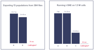
CellEngine is orders of magnitude faster than other analysis software. Optimized by performance experts to achieve the speed needed to analyze large, high-dimensional datasets, CellEngine provides rapid analysis that is easy for anyone to use—no coding skills required. CellEngine can run advanced analysis pipelines with PhenoGraph, t-SNE, UMAP, SOM, and more on millions of cells in just minutes. In a head-to-head comparison of CellEngine and other software, 384 data files were analyzed from 13 different populations. The other software took hours, while CellEngine took seconds. With a more complex t-SNE visualization, CellEngine only took minutes instead of hours.
CellEngine™: Transitioning to the Next Generation of Cytometry Analysis Software

Versatile analysis and advanced visualization of high-dimensional cytometry data

A wide variety of options for visualizations can be created within CellEngine, all driven by your experiment metadata. Dot plots, histograms, heatmaps, dose response curves, metadata-driven layouts, and algorithmic visualizations are available to distill your data into figures and slides.

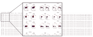
CellEngine can easily create pivot tables and batched analysis across samples or experimental conditions, allowing quick visualization of large datasets. Metadata-driven layouts take seconds to create by simply annotating files and selecting visualization parameters. In the pivot table below, various samples are organized by time points, conditions, and patient, providing a highly informative output for longitudinal studies.
CellEngine™: Transitioning to the Next Generation of Cytometry Analysis Software

Supervised autogating for trusted data in less time

CellEngine includes a built-in tool for supervised autogating by using the power of machine-learning to automatically tailor your gates. The tool uses a small set of manually gated files from your dataset and adjusts gate positions to match in seconds. This approach reduces subjectivity and increases consistency, while saving massive amounts of time.

How does CellCarta utilize CellEngine to meet our clients’ needs?

As a flow and mass cytometry CRO, CellCarta generates many high-dimensional data files daily and outgrew our previous software. CellEngine’s speed and visualization features are invaluable for analyzing multi-year clinical studies with thousands of samples. Because CellEngine is cloud-based, it allows for secure, easy communication of data and analysis to our clients. Its 21 CFR 11 compliance allows us to use it in primary and secondary endpoint studies. CellEngine’s rich API integrates into our bioinformatics pipelines, performing tasks such as automatic data upload and pre-processing. CellCarta provides more informative data to our clients, faster, helping them to rapidly advance their therapeutics.

Welcome to a new generation of cytometry analysis software.  Let CellEngine make your data talk. Sign up for a free two-month trial or contact us for a live demo.

Watch the following video from our expert to learn more.


About the author:

author photo

Susan Reynolds is a Scientific Business Director at CellCarta specializing in multi-omic flow cytometric application platforms. She has held various roles in the industry with emphasis on strategic development and commercialization of emerging novel platforms.


CellEngine: Get The Next-Generation Cytometry Analysis Software

September 12, 2022

CellEngine™: Transitioning to the Next Generation of Cytometry Analysis Software

Today’s high-dimensional cytometry can measure dozens of analytes for millions of cells, and high-throughput cytometers can generate thousands of files, totaling hundreds of gigabytes of data. With most software, analyzing this amount of data is slow, creating complex visualizations can be difficult or impossible, and sharing data with coworkers and collaborators can be cumbersome. Finally, few programs provide regulatory compliance features necessary for running clinical trials.

CellEngineTM addresses the gaps where other software fails

CellEngine is a cloud-based cytometry analysis software platform, designed to tackle these challenges. CellEngine provides fast, full analytical and visualization functionality in one software program. Its cloud-native design allows easy collaboration, while providing enterprise-grade data safety and security. A first-class API offers full access to any CellEngine feature, allowing easy integration into bioinformatics pipelines and LIMS systems. For research performed in regulated environments, CellEngine supplies all the features needed for use in 21 CFR 11-compliant workflows.

Lightning-fast analysis of large, complex datasets

CellEngine is orders of magnitude faster than other analysis software. Optimized by performance experts to achieve the speed needed to analyze large, high-dimensional datasets, CellEngine provides rapid analysis that is easy for anyone to use—no coding skills required. CellEngine can run advanced analysis pipelines with PhenoGraph, t-SNE, UMAP, SOM, and more on millions of cells in just minutes. In a head-to-head comparison of CellEngine and other software, 384 data files were analyzed from 13 different populations. The other software took hours, while CellEngine took seconds. With a more complex t-SNE visualization, CellEngine only took minutes instead of hours.

CellEngine™: Transitioning to the Next Generation of Cytometry Analysis Software

Versatile analysis and advanced visualization of high-dimensional cytometry data

A wide variety of options for visualizations can be created within CellEngine, all driven by your experiment metadata. Dot plots, histograms, heatmaps, dose response curves, metadata-driven layouts, and algorithmic visualizations are available to distill your data into figures and slides.

CellEngine can easily create pivot tables and batched analysis across samples or experimental conditions, allowing quick visualization of large datasets. Metadata-driven layouts take seconds to create by simply annotating files and selecting visualization parameters. In the pivot table below, various samples are organized by time points, conditions, and patient, providing a highly informative output for longitudinal studies.

CellEngine™: Transitioning to the Next Generation of Cytometry Analysis Software

Supervised autogating for trusted data in less time

CellEngine includes a built-in tool for supervised autogating by using the power of machine-learning to automatically tailor your gates. The tool uses a small set of manually gated files from your dataset and adjusts gate positions to match in seconds. This approach reduces subjectivity and increases consistency, while saving massive amounts of time.

How does CellCarta utilize CellEngine to meet our clients’ needs?

As a flow and mass cytometry CRO, CellCarta generates many high-dimensional data files daily and outgrew our previous software. CellEngine’s speed and visualization features are invaluable for analyzing multi-year clinical studies with thousands of samples. Because CellEngine is cloud-based, it allows for secure, easy communication of data and analysis to our clients. Its 21 CFR 11 compliance allows us to use it in primary and secondary endpoint studies. CellEngine’s rich API integrates into our bioinformatics pipelines, performing tasks such as automatic data upload and pre-processing. CellCarta provides more informative data to our clients, faster, helping them to rapidly advance their therapeutics.

Welcome to a new generation of cytometry analysis software.  Let CellEngine make your data talk. Sign up for a free two-month trial or contact us for a live demo.

Watch the following video from our expert to learn more.


About the author:

author photo

Susan Reynolds is a Scientific Business Director at CellCarta specializing in multi-omic flow cytometric application platforms. She has held various roles in the industry with emphasis on strategic development and commercialization of emerging novel platforms.

HER2-Low Breast Cancer: New Insights from Enhertu Clinical Trials

June 23, 2022

CellTalk_HER2-MRM

Recent results presented during the keynote address at the 2022 American Society of Clinical Oncology (ASCO) annual meeting highlighted the emergence of a new era for breast cancer treatment. The landmark study on the HER2-directed therapy, Enhertu, found that patients with HER2-negative breast cancer may soon have the opportunity to receive this new treatment based on exceptional outcomes in a Phase 3 clinical trial.

In this pivotal trial, patients with HER2-low metastatic breast cancer treated with Enhertu had a 49% lower risk of disease progression or death vs. patients treated with chemotherapy alone.1, 2 In most subgroups the progression free survival (PFS) was nearly doubled in the Enhertu arm vs. chemotherapy. HER2-positive tumors are routinely identified by pathologists using a combination of immunohistochemistry (IHC) tests and in situ hybridization (ISH). However, the trial results clearly demonstrated that  HER2-low tumors (HER 1+ and 2+) also need clear identification, especially since they are more difficult to distinguish from HER2-negative with traditional methods. 2

Meeting the need for a more quantitative methodology

The Enhertu trial has identified a clear unmet need for more quantitative methodologies for HER2 detection, specifically the ability to distinguish low HER2, and potentially even “ultra low-HER2”, expressing populations, who will likely benefit from Enhertu treatment, from those that are truly negative.

To overcome the challenges of antibody-based IHC in detecting HER2-low metastatic breast cancer, CellCarta has developed a precision measurement approach leveraging mass spectrometry based Multiple Reaction Monitoring (MRM). The MRM platform has exceptional sensitivity, specificity, and quantitative precision to measure protein analytes, providing one of the most specific assays for protein measurement.

When it comes to measuring HER2, CellCarta has demonstrated that MRM can provide valuable information to quantitatively characterize HER2 expression in a patient’s tumor biopsy. Our method has the potential to improve patient selection and ultimately expand the population of patients who will likely benefit from novel therapies such as Enhertu.

Exceptional specificity for detection of single or multiplexed cancer protein biomarkers

MRM offers absolute quantification and even provides accurate multiplexed quantitation of clinically relevant biomarkers such as HER2. CellCarta offers off-the-shelf multiplexed panels, including our MRM assay for HER2, or rapid development and validation of custom assays, depending on your study needs. We design and validate multiplexes of cancer protein biomarker panels of up to 15 proteins. All of our assays are validated for clinical use under CAP/CLIA and GCLP environments.


About the author:

author photo

Nick Dupuis is a Senior Director of Scientific Business Development at CellCarta, specialized in proteomics and biophysical measurement. He has held various roles in industry including as a scientist in LDT/IVD product and applications development, and business development for emerging analytical platforms.

References

  1. Modi S, Jacot W, Yamashita T, et al. Trastuzumab Deruxtecan in Previously Treated HER2-Low Advanced Breast Cancer. NEJM 2022;doi:10.1056/NEJMoa2203690.
  2. Liu A. Fierce Biotech. ASCO: HER2 diagnostics need a revolution as AstraZeneca, Daiichi’s Enhertu looks to redefine breast cancer. June 13, 2022. https://www.fiercebiotech.com/medtech/asco-her2-diagnostics-need-revolution-astrazeneca-daiichis-enhertu-looks-redefine-breast

HER2-Low Breast Cancer: New Insights from Enhertu Clinical Trials

June 23, 2022

CellTalk_HER2-MRM

Recent results presented during the keynote address at the 2022 American Society of Clinical Oncology (ASCO) annual meeting highlighted the emergence of a new era for breast cancer treatment. The landmark study on the HER2-directed therapy, Enhertu, found that patients with HER2-negative breast cancer may soon have the opportunity to receive this new treatment based on exceptional outcomes in a Phase 3 clinical trial.

In this pivotal trial, patients with HER2-low metastatic breast cancer treated with Enhertu had a 49% lower risk of disease progression or death vs. patients treated with chemotherapy alone.1, 2 In most subgroups the progression free survival (PFS) was nearly doubled in the Enhertu arm vs. chemotherapy. HER2-positive tumors are routinely identified by pathologists using a combination of immunohistochemistry (IHC) tests and in situ hybridization (ISH). However, the trial results clearly demonstrated that  HER2-low tumors (HER 1+ and 2+) also need clear identification, especially since they are more difficult to distinguish from HER2-negative with traditional methods. 2

Meeting the need for a more quantitative methodology

The Enhertu trial has identified a clear unmet need for more quantitative methodologies for HER2 detection, specifically the ability to distinguish low HER2, and potentially even “ultra low-HER2”, expressing populations, who will likely benefit from Enhertu treatment, from those that are truly negative.

To overcome the challenges of antibody-based IHC in detecting HER2-low metastatic breast cancer, CellCarta has developed a precision measurement approach leveraging mass spectrometry based Multiple Reaction Monitoring (MRM). The MRM platform has exceptional sensitivity, specificity, and quantitative precision to measure protein analytes, providing one of the most specific assays for protein measurement.

When it comes to measuring HER2, CellCarta has demonstrated that MRM can provide valuable information to quantitatively characterize HER2 expression in a patient’s tumor biopsy. Our method has the potential to improve patient selection and ultimately expand the population of patients who will likely benefit from novel therapies such as Enhertu.

Exceptional specificity for detection of single or multiplexed cancer protein biomarkers

MRM offers absolute quantification and even provides accurate multiplexed quantitation of clinically relevant biomarkers such as HER2. CellCarta offers off-the-shelf multiplexed panels, including our MRM assay for HER2, or rapid development and validation of custom assays, depending on your study needs. We design and validate multiplexes of cancer protein biomarker panels of up to 15 proteins. All of our assays are validated for clinical use under CAP/CLIA and GCLP environments.


About the author:

author photo

Nick Dupuis is a Senior Director of Scientific Business Development at CellCarta, specialized in proteomics and biophysical measurement. He has held various roles in industry including as a scientist in LDT/IVD product and applications development, and business development for emerging analytical platforms.

References

  1. Modi S, Jacot W, Yamashita T, et al. Trastuzumab Deruxtecan in Previously Treated HER2-Low Advanced Breast Cancer. NEJM 2022;doi:10.1056/NEJMoa2203690.
  2. Liu A. Fierce Biotech. ASCO: HER2 diagnostics need a revolution as AstraZeneca, Daiichi’s Enhertu looks to redefine breast cancer. June 13, 2022. https://www.fiercebiotech.com/medtech/asco-her2-diagnostics-need-revolution-astrazeneca-daiichis-enhertu-looks-redefine-breast

Effective Biomarker Strategies for Emerging Multiple Myeloma Therapies

June 9, 2021

Section image

Targeting BMCA in multiple myeloma (MM) has leapt into clinical practice, most recently with the approvals of a new antibody drug conjugate (BLENREP, GSK, 2020) and a chimeric antigen receptor T-cell therapy (CAR T, ABECMA, BMS, 2021). While BCMA has shown great promise, several new drugs directed at other targets for treatment of MM have recently been approved or are in development, greatly expanding treatment options in relapsed or refractory settings.

While there is an increasing number of clinical trials investigating drugs targeting BCMA (from 5 in 2015 to >30 in 2019 according to clinicaltrials.gov), other targets have been pursued, including: CD38, HDAC, and SLAMF7.

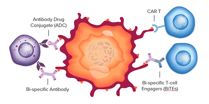
For each of these targets, multiple therapeutic modalities across small molecules, monoclonal antibodies (mAb), antibody drug conjugates (ADCs), bi-specific antibodies and more recently CD3-directed bi-specifics T-cell engagers (BiTEs) and CAR-T therapies are being investigated. However, each target and drug combination represent a unique translational biomarker challenge with different combinations of mechanisms of action (MOA) and therapeutic pathways. This complexity is something we are keenly focused on at CellCarta where we bring a comprehensive technology approach to addressing biomarker strategies and deliver biomarker data to support clinical programs.

Section image

Biomarkers to stratify MM patients with greater precision

Patient stratification in clinical trials depends on prognostic factors such as disease burden and staging, tumor biology, but also on the expression of the target proteins of the investigational treatments. To enable stratification with greater precision, CellCarta offers multiple assays investigating key biomarkers in CD138 enriched bone marrow cells.

Using bone marrow biopsies, IHC can be performed to further profile the tumor as well as the tumor micro-environment (TME) using key markers such as CD38, CD138, BCMA.

Monitoring immune responses in MM patients using key biomarkers

Flow cytometry allows the direct measurement of cells with abnormal phenotypes associated with disease progression. At CellCarta we deploy off-the-shelf and custom multi-color flow panels to phenotypically characterize a variety of immune subsets acting as key biomarkers across all phases of drug development.

A specific plasma cell panel, including markers such as CD138, CD38, BCMA and PD-L1, was developed to monitor the presence of abnormal plasma cells in both blood and bone marrow aspirate clinical samples. BD TrucountTM tubes can provide both frequencies and absolute counts readouts. Using this panel, important prognostic factors relating to treatments such as BCMA and PD-L1 can be investigated on subsets of interest. For individuals undergoing anti-CD38 therapy, the use of a polyclonal anti-CD38 antibody allows us to detect the receptor. The tumor microenvironment (TME) can also be investigated using flow cytometry and customized panels.

Tracking the course of MM with soluble biomarkers

Soluble blood-based biomarkers have been used to diagnose and monitor the course of MM since the inclusion of serum free light chain (FLC) concentrations in the 2014 IMWG criteria. While FLC assays are a routine part of the MM clinical biomarker analysis, along with the haemato-pathological and flow cytometry workflows, the emergence of BCMA targeted therapies has led to the addition of soluble BCMA concentrations as a critical biomarker. At CellCarta we have developed an off-the-shelf hybrid IP-LC-MRM based assay for the quantification of soluble BCMA in plasma for both diagnostic and prognostic utility. The assay is notably robust to interference from endogenous ligands (APRIL and BAFF), and therapeutic antibodies.

These biomarker strategies are complemented with broader immune monitoring capabilities essential in investigating a patient’s response to treatments. These include cytokine measurement by Meso Scale Discovery (CLIA-validated proinflammatory panel) and broad immune monitoring by flow cytometry. CellCarta’s deep expertise in immunology helps us address all the needs when it comes to MM clinical research.


About the author:

author photo

Nick Dupuis is a Scientific Business Director at CellCarta, specialized in proteomics and biophysical measurement. He has held various roles in industry including as a scientist in LDT/IVD product development and supporting applications and business development for emerging analytical platforms.

author photo

Damien Montamat-Sicotte is a Scientific Business Director at CellCarta, specializing in the flow Cytometry platform. With a PhD in immunology and post-doctoral expertise from various institutions, Damien has profuse experience in managing the processing and analysis of clinical samples by flow cytometry in an immune monitoring context.

Effective Biomarker Strategies for Emerging Multiple Myeloma Therapies

June 9, 2021

Section image

Targeting BMCA in multiple myeloma (MM) has leapt into clinical practice, most recently with the approvals of a new antibody drug conjugate (BLENREP, GSK, 2020) and a chimeric antigen receptor T-cell therapy (CAR T, ABECMA, BMS, 2021). While BCMA has shown great promise, several new drugs directed at other targets for treatment of MM have recently been approved or are in development, greatly expanding treatment options in relapsed or refractory settings.

While there is an increasing number of clinical trials investigating drugs targeting BCMA (from 5 in 2015 to >30 in 2019 according to clinicaltrials.gov), other targets have been pursued, including: CD38, HDAC, and SLAMF7.

For each of these targets, multiple therapeutic modalities across small molecules, monoclonal antibodies (mAb), antibody drug conjugates (ADCs), bi-specific antibodies and more recently CD3-directed bi-specifics T-cell engagers (BiTEs) and CAR-T therapies are being investigated. However, each target and drug combination represent a unique translational biomarker challenge with different combinations of mechanisms of action (MOA) and therapeutic pathways. This complexity is something we are keenly focused on at CellCarta where we bring a comprehensive technology approach to addressing biomarker strategies and deliver biomarker data to support clinical programs.

Section image

Biomarkers to stratify MM patients with greater precision

Patient stratification in clinical trials depends on prognostic factors such as disease burden and staging, tumor biology, but also on the expression of the target proteins of the investigational treatments. To enable stratification with greater precision, CellCarta offers multiple assays investigating key biomarkers in CD138 enriched bone marrow cells.

Using bone marrow biopsies, IHC can be performed to further profile the tumor as well as the tumor micro-environment (TME) using key markers such as CD38, CD138, BCMA.

Monitoring immune responses in MM patients using key biomarkers

Flow cytometry allows the direct measurement of cells with abnormal phenotypes associated with disease progression. At CellCarta we deploy off-the-shelf and custom multi-color flow panels to phenotypically characterize a variety of immune subsets acting as key biomarkers across all phases of drug development.

A specific plasma cell panel, including markers such as CD138, CD38, BCMA and PD-L1, was developed to monitor the presence of abnormal plasma cells in both blood and bone marrow aspirate clinical samples. BD TrucountTM tubes can provide both frequencies and absolute counts readouts. Using this panel, important prognostic factors relating to treatments such as BCMA and PD-L1 can be investigated on subsets of interest. For individuals undergoing anti-CD38 therapy, the use of a polyclonal anti-CD38 antibody allows us to detect the receptor. The tumor microenvironment (TME) can also be investigated using flow cytometry and customized panels.

Tracking the course of MM with soluble biomarkers

Soluble blood-based biomarkers have been used to diagnose and monitor the course of MM since the inclusion of serum free light chain (FLC) concentrations in the 2014 IMWG criteria. While FLC assays are a routine part of the MM clinical biomarker analysis, along with the haemato-pathological and flow cytometry workflows, the emergence of BCMA targeted therapies has led to the addition of soluble BCMA concentrations as a critical biomarker. At CellCarta we have developed an off-the-shelf hybrid IP-LC-MRM based assay for the quantification of soluble BCMA in plasma for both diagnostic and prognostic utility. The assay is notably robust to interference from endogenous ligands (APRIL and BAFF), and therapeutic antibodies.

These biomarker strategies are complemented with broader immune monitoring capabilities essential in investigating a patient’s response to treatments. These include cytokine measurement by Meso Scale Discovery (CLIA-validated proinflammatory panel) and broad immune monitoring by flow cytometry. CellCarta’s deep expertise in immunology helps us address all the needs when it comes to MM clinical research.


About the author:

author photo

Nick Dupuis is a Scientific Business Director at CellCarta, specialized in proteomics and biophysical measurement. He has held various roles in industry including as a scientist in LDT/IVD product development and supporting applications and business development for emerging analytical platforms.

author photo

Damien Montamat-Sicotte is a Scientific Business Director at CellCarta, specializing in the flow Cytometry platform. With a PhD in immunology and post-doctoral expertise from various institutions, Damien has profuse experience in managing the processing and analysis of clinical samples by flow cytometry in an immune monitoring context.

Steps for Developing Multiplex IHC Assays: Comprehensive Guide

May 26, 2021

Section image

Multiplex immunohistochemistry (mIHC) enables the identification of different immune cell subsets, their activation state, and their spatial distribution in the tumor microenvironment (TME). The multiplex platform allows maximal data collection from tissue sparing clinical samples.

Simultaneous detection of multiple protein biomarkers on a single tissue section can come with challenges. Therefore, in a clinical setting, a rigorous optimization and validation process must be put in place to ensure the assay can be trusted.

Here we will discuss the optimization and validation process we follow at CellCarta for a chromogenic mIHC assay.

Step 1

Optimizing the single IHC of each target. Identify monoclonal antibody clone, antigen retrieval conditions and antibody incubation times for each target using positive control tissues.

Step 2

Assembling the multiplex panel. The order of antibody target and chromogen deposition is determined.

Step 3

Testing the optimized staining conditions in the multiplex protocol. Calibration experiments are performed to ensure that for each target, the signal is specific and balanced across the multiplex panel.

Step 4

Comparing the mIHC assay to its respective validated single plex IHC counterparts. Serial slides stained for each validated single plex IHC are compared to the multiplex staining.

Step 5

Performing a precision test. As a final stage, serial slides are sequentially stained with the antibodies or an IgG isotype matched control (negative control).

We use digital image analysis methodologies to do in-depth immune profiling of the TME. These methods can complement a pathologist analysis by providing a detailed quantitative analysis. Image analysis tools are very useful for the development of biomarker strategies.

Read our white paper for details on the development of our triplex chromogenic mIHC CD8/GZMB/FOXP3:


About the Author:

author photo

Susan McCarthy is a scientific business director at CellCarta, dedicated to the histopathology platform. With over 18 years of experience in biomarker and clinical assay development, including at the Moffitt Cancer Center, Susan has a strong background in immuno-oncology, molecular biology, and digital pathology.

Steps for Developing Multiplex IHC Assays: Comprehensive Guide

May 26, 2021

Section image

Multiplex immunohistochemistry (mIHC) enables the identification of different immune cell subsets, their activation state, and their spatial distribution in the tumor microenvironment (TME). The multiplex platform allows maximal data collection from tissue sparing clinical samples.

Simultaneous detection of multiple protein biomarkers on a single tissue section can come with challenges. Therefore, in a clinical setting, a rigorous optimization and validation process must be put in place to ensure the assay can be trusted.

Here we will discuss the optimization and validation process we follow at CellCarta for a chromogenic mIHC assay.

Step 1

Optimizing the single IHC of each target. Identify monoclonal antibody clone, antigen retrieval conditions and antibody incubation times for each target using positive control tissues.

Step 2

Assembling the multiplex panel. The order of antibody target and chromogen deposition is determined.

Step 3

Testing the optimized staining conditions in the multiplex protocol. Calibration experiments are performed to ensure that for each target, the signal is specific and balanced across the multiplex panel.

Step 4

Comparing the mIHC assay to its respective validated single plex IHC counterparts. Serial slides stained for each validated single plex IHC are compared to the multiplex staining.

Step 5

Performing a precision test. As a final stage, serial slides are sequentially stained with the antibodies or an IgG isotype matched control (negative control).

We use digital image analysis methodologies to do in-depth immune profiling of the TME. These methods can complement a pathologist analysis by providing a detailed quantitative analysis. Image analysis tools are very useful for the development of biomarker strategies.

Read our white paper for details on the development of our triplex chromogenic mIHC CD8/GZMB/FOXP3:


About the Author:

author photo

Susan McCarthy is a scientific business director at CellCarta, dedicated to the histopathology platform. With over 18 years of experience in biomarker and clinical assay development, including at the Moffitt Cancer Center, Susan has a strong background in immuno-oncology, molecular biology, and digital pathology.

MHC Peptide Analysis: Insights from Eustache Paramithiotis

July 13, 2020

Discussing MHC Peptide Analysis with Eustache Paramithiotis

We recently sat down with our Vice-President of Research and Development, Eustache Paramithiotis to discuss some of the finer points of MHC peptide analysis, why it is important for a complete understanding of an immune response, and how it is supporting new waves in drug development.

Q: Let’s begin with the basics, what is the MHC system and how does it help regulate immune responses?

A: The immune system surveys what is going on in the body through the MHC. There are three kinds of MHC, Class I, Class II, and non-classical, all presenting peptides and sometimes other things like lipids. This is how T cells do their surveillance and identify anything unusual or “non-self”. Thus, the MHC is essential for launching an immune response, the first step of it all!

Q: With the recent advancement of immune therapies, is MHC peptide presentation critical information to know?

A: In fact, it is absolutely essential! Modern immune therapies involve the host immune system. The antigen is either directly provided to educate the immune cells, or modified cells, as per CAR-T cell therapy, are introduced to attack the tumor. Some immune responses are obtainable with non-MHC based system, including innate immunity and NK cells, but an optimal and complete immune response will require MHC presentation.

Q: Characterizing MHC peptides is a significant technical challenge, how does your team tackle it?

A: Immunologists had determined many years ago that antigen was presented in the ‘context of MHC’, a concept developed from functional studies, though they didn’t know how that actually worked. The concept was confirmed by crystal structures, which showed directly that antigen – in this case a peptide – was embedded in the MHC and the MHC-peptide complex was what the T cell receptors bound to. The peptide’s length and composition may vary with the type of MHC, but the concept remains the same, a peptide embedded in a macromolecule and both presented together. All the analytical techniques that have been developed over time to isolate, process, and analyze peptides come into play here. Having the capability to perform accurate and reproducible mass spectrometry and evaluate results with strong bioinformatic tools is essential for productive experiments. All of which we invested in and can now do at scale.

Q: Can you expand on the importance of the direct measurement of peptides?

A: The most physiologically relevant way to see what the host is presenting is to look directly in the tissue. Given the structure of the MHC Class I or Class II, presented peptides tend to have themes in their sequence, anchor points and motifs, which led to the use of computer algorithms to predict what would be presented. With all the projects we have done, we have seen that what is actually presented is far more complex than what the algorithms are able to tell us. The algorithms are much better at modelling peptide-receptor interactions once you have a peptide sequence, but since we don’t fully understand the regulation of antigen processing and selection for what will get loaded onto the MHC it is difficult to accurately predict what will get presented.

So, when it comes to the kind of personalized targets that are ideal for cancer immunotherapies, including neoepitopes (peptides with somatic mutations), prediction is inefficient. The variety of modifications we can observe directly in tissues is much broader than the one obtained with prediction algorithms. Nowadays, if you cannot directly observe what is being presented, there will be gaps in your understanding.

Q: In addition to direct detection, what are some of the learnings about MHC peptides that differentiates Caprion-HistoGeneX’s approach from what others are doing?

A: MHC presentation is necessary to initiate and maintain an immune response, but it’s not the only thing you need. To be able to judge how well peptides induce an immune response, you need a range of immune monitoring or immune evaluation capabilities. That is where Caprion-HistoGeneX comes in and has put all those pieces together. We provide the complete picture of all the factors involved, from identifying the target in tissue, to the levels of MHC I expression, and to characterizing the responsiveness of the host T cells to target.

Q: How does the Antigen Atlas fit into this picture and your ability to characterize the MHC presentome?

A: The Antigen Atlas is primarily a prioritization tool. It was built to help prioritize the targets based on their lack of presentation in other tissues. The Atlas contains MHC I presented peptides from a library of healthy and tumor adjacent tissues (usually a bit more inflamed than healthy tissue), and it includes a wide range of different MHC I alleles. The database has on the order of 400,000 entries right now and is often updated to stay current. If you have a peptide you are interested in from a target tissue, you can check if it is presented in other tissues. Typically, therapies are concerned with understanding on-target and off-target effects. Immune therapies as well, but also need to understand on-target but off-tissue effects, because you don’t want to trigger an attack to the liver 5 years after curing a lung cancer. A large database, made to our quality standards, can also do a lot of other things than just help prioritize targets. For example, you can use it to better understand baseline mutation presentation in healthy tissues, identify protein presentation hotspots that are independent of the MHC I alleles an individual expresses, and identify tissue-specific presentation patterns to name a few.

Q: In the next few years, what innovations do you think MHC peptide analysis will help drive?

A: Right now, all of it is focused on MHC Class I and effector/cytotoxic T cells, because that is the pointy end of the stick. To mount a complete robust immune response with memory, you need Class II involvement and that has not yet become as prominent in the current analysis. We will eventually need to know what helper and regulatory T cells as well as B cell are presenting, which means both Class I and Class II MHC. Another aspect we will need to further investigate is the heterogeneity of presentation. Right now, we don’t really know where in the tissue peptides are presented. Are they presented in a homogenous way, or are they presented in pockets, which may be less accessible to infiltrating lymphocytes? A better understanding of peptide distribution, especially when it comes to solid tumor therapies, is going to become very important.

Q: What are your key takeaways or what you would like the readers to know about MHC peptide analysis with Caprion-HistoGeneX?

A: Key takeaways would be that MHC peptide analysis is now practical, it’s doable on just about any kind of system. It is also necessary for the understanding of immune responses. And finally, it should be considered as an essential part of a larger assessment of immune function. Caprion-HistoGeneX can evaluate, understand, and monitor the entire immune response. You get to find the antigens and what is presented, but then we’ll also help you figure out what to do with them and how to get them to react.

MHC Peptide Analysis: Insights from Eustache Paramithiotis

July 13, 2020

Discussing MHC Peptide Analysis with Eustache Paramithiotis

We recently sat down with our Vice-President of Research and Development, Eustache Paramithiotis to discuss some of the finer points of MHC peptide analysis, why it is important for a complete understanding of an immune response, and how it is supporting new waves in drug development.

Q: Let’s begin with the basics, what is the MHC system and how does it help regulate immune responses?

A: The immune system surveys what is going on in the body through the MHC. There are three kinds of MHC, Class I, Class II, and non-classical, all presenting peptides and sometimes other things like lipids. This is how T cells do their surveillance and identify anything unusual or “non-self”. Thus, the MHC is essential for launching an immune response, the first step of it all!

Q: With the recent advancement of immune therapies, is MHC peptide presentation critical information to know?

A: In fact, it is absolutely essential! Modern immune therapies involve the host immune system. The antigen is either directly provided to educate the immune cells, or modified cells, as per CAR-T cell therapy, are introduced to attack the tumor. Some immune responses are obtainable with non-MHC based system, including innate immunity and NK cells, but an optimal and complete immune response will require MHC presentation.

Q: Characterizing MHC peptides is a significant technical challenge, how does your team tackle it?

A: Immunologists had determined many years ago that antigen was presented in the ‘context of MHC’, a concept developed from functional studies, though they didn’t know how that actually worked. The concept was confirmed by crystal structures, which showed directly that antigen – in this case a peptide – was embedded in the MHC and the MHC-peptide complex was what the T cell receptors bound to. The peptide’s length and composition may vary with the type of MHC, but the concept remains the same, a peptide embedded in a macromolecule and both presented together. All the analytical techniques that have been developed over time to isolate, process, and analyze peptides come into play here. Having the capability to perform accurate and reproducible mass spectrometry and evaluate results with strong bioinformatic tools is essential for productive experiments. All of which we invested in and can now do at scale.

Q: Can you expand on the importance of the direct measurement of peptides?

A: The most physiologically relevant way to see what the host is presenting is to look directly in the tissue. Given the structure of the MHC Class I or Class II, presented peptides tend to have themes in their sequence, anchor points and motifs, which led to the use of computer algorithms to predict what would be presented. With all the projects we have done, we have seen that what is actually presented is far more complex than what the algorithms are able to tell us. The algorithms are much better at modelling peptide-receptor interactions once you have a peptide sequence, but since we don’t fully understand the regulation of antigen processing and selection for what will get loaded onto the MHC it is difficult to accurately predict what will get presented.

So, when it comes to the kind of personalized targets that are ideal for cancer immunotherapies, including neoepitopes (peptides with somatic mutations), prediction is inefficient. The variety of modifications we can observe directly in tissues is much broader than the one obtained with prediction algorithms. Nowadays, if you cannot directly observe what is being presented, there will be gaps in your understanding.

Q: In addition to direct detection, what are some of the learnings about MHC peptides that differentiates Caprion-HistoGeneX’s approach from what others are doing?

A: MHC presentation is necessary to initiate and maintain an immune response, but it’s not the only thing you need. To be able to judge how well peptides induce an immune response, you need a range of immune monitoring or immune evaluation capabilities. That is where Caprion-HistoGeneX comes in and has put all those pieces together. We provide the complete picture of all the factors involved, from identifying the target in tissue, to the levels of MHC I expression, and to characterizing the responsiveness of the host T cells to target.

Q: How does the Antigen Atlas fit into this picture and your ability to characterize the MHC presentome?

A: The Antigen Atlas is primarily a prioritization tool. It was built to help prioritize the targets based on their lack of presentation in other tissues. The Atlas contains MHC I presented peptides from a library of healthy and tumor adjacent tissues (usually a bit more inflamed than healthy tissue), and it includes a wide range of different MHC I alleles. The database has on the order of 400,000 entries right now and is often updated to stay current. If you have a peptide you are interested in from a target tissue, you can check if it is presented in other tissues. Typically, therapies are concerned with understanding on-target and off-target effects. Immune therapies as well, but also need to understand on-target but off-tissue effects, because you don’t want to trigger an attack to the liver 5 years after curing a lung cancer. A large database, made to our quality standards, can also do a lot of other things than just help prioritize targets. For example, you can use it to better understand baseline mutation presentation in healthy tissues, identify protein presentation hotspots that are independent of the MHC I alleles an individual expresses, and identify tissue-specific presentation patterns to name a few.

Q: In the next few years, what innovations do you think MHC peptide analysis will help drive?

A: Right now, all of it is focused on MHC Class I and effector/cytotoxic T cells, because that is the pointy end of the stick. To mount a complete robust immune response with memory, you need Class II involvement and that has not yet become as prominent in the current analysis. We will eventually need to know what helper and regulatory T cells as well as B cell are presenting, which means both Class I and Class II MHC. Another aspect we will need to further investigate is the heterogeneity of presentation. Right now, we don’t really know where in the tissue peptides are presented. Are they presented in a homogenous way, or are they presented in pockets, which may be less accessible to infiltrating lymphocytes? A better understanding of peptide distribution, especially when it comes to solid tumor therapies, is going to become very important.

Q: What are your key takeaways or what you would like the readers to know about MHC peptide analysis with Caprion-HistoGeneX?

A: Key takeaways would be that MHC peptide analysis is now practical, it’s doable on just about any kind of system. It is also necessary for the understanding of immune responses. And finally, it should be considered as an essential part of a larger assessment of immune function. Caprion-HistoGeneX can evaluate, understand, and monitor the entire immune response. You get to find the antigens and what is presented, but then we’ll also help you figure out what to do with them and how to get them to react.