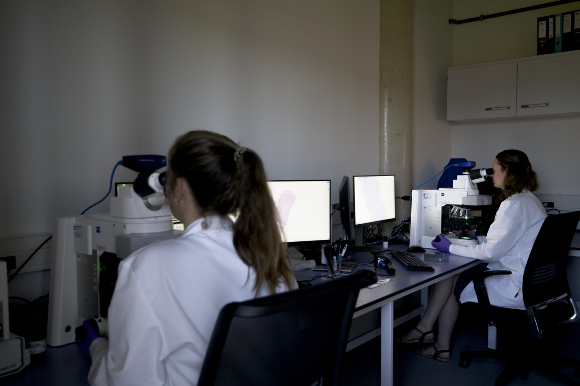
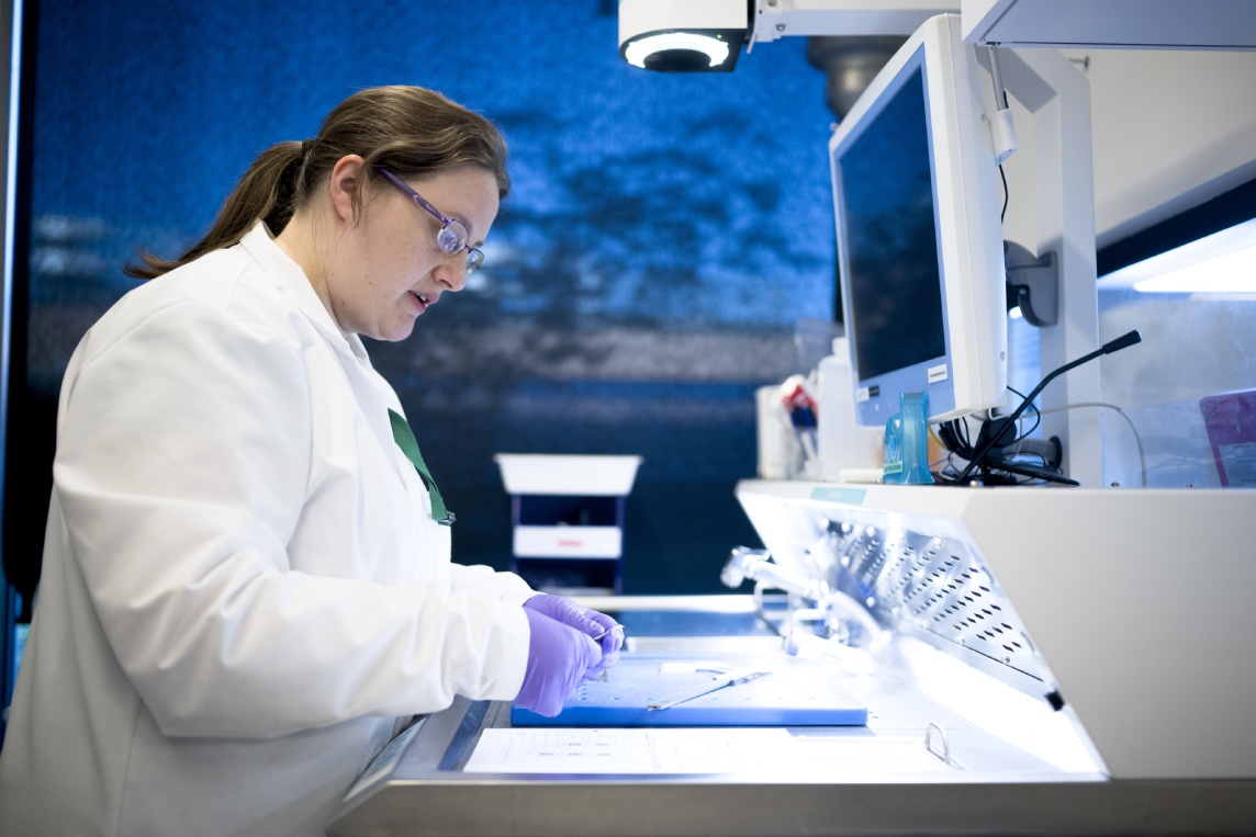
From singleplex chromogenic to multiplex IHC assays, we deliver high quality, reproducible staining and imaging services for implementation in clinical trials globally.
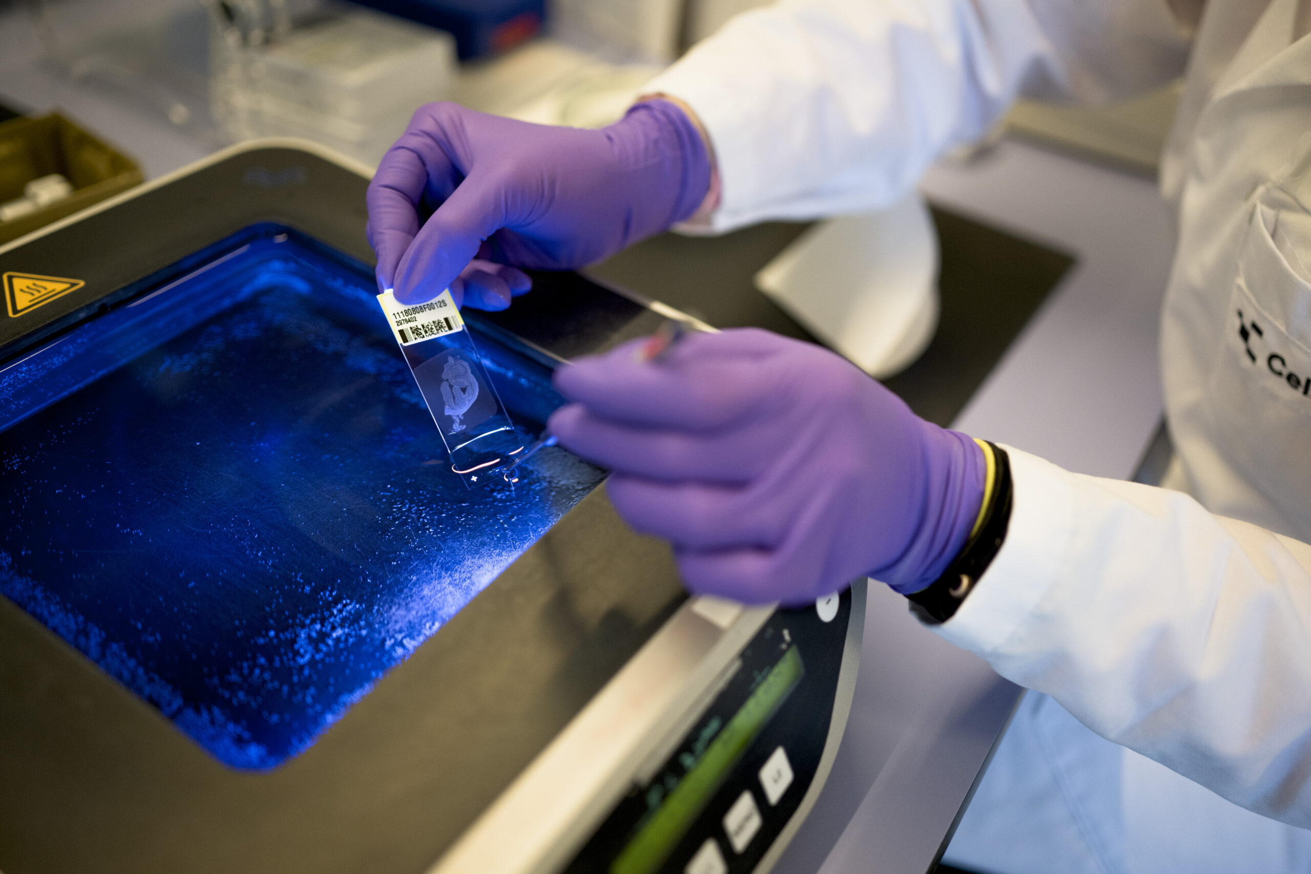
CellCarta’s leading staining and imaging platforms support histopathology services for the entire therapeutic development process.
Our platforms and protocols are cross-validated by our histopathology laboratories in the USA, Europe, and China, ensuring harmonized, reliable results regardless of your study’s location.
We take an agnostic approach to assay development and clinical testing, delivering services across a large suite of staining instruments.
Unsure which platform is best suited for your study? Our histopathology team can help inform your selection.
We offer a multitude of brightfield and fluorescent scanners for cell imaging, including:
Our imaging platforms have been implemented in over 1000 clinical trials, and can support your study at the exploratory, primary, and secondary endpoint.
We can perform hematoxylin and eosin (H&E) staining on your clinical samples, allowing you to investigate therapeutic efficacy, including tissue inflammation, tumor-infiltrating lymphocytes (TILs), and tertiary lymphoid structures (TLS).
Our H&E staining services can visualize cellular and tissue structures in FFPE, fresh, and frozen tissue.
All H&E scoring and annotation is performed by our in-house pathologists.
Our pathologists and imaging scientists have expertise implementing AI-supported digital image analysis platforms for dataset evaluation, including:
Our pathologists perform rigorous review and QC of all datasets, ensuring your study’s results are high-quality and reproducible.
We take an agnostic approach to assay development and clinical testing, delivering services across a large suite of staining instruments.
Unsure which platform is best suited for your study? Our histopathology team can help inform your selection.
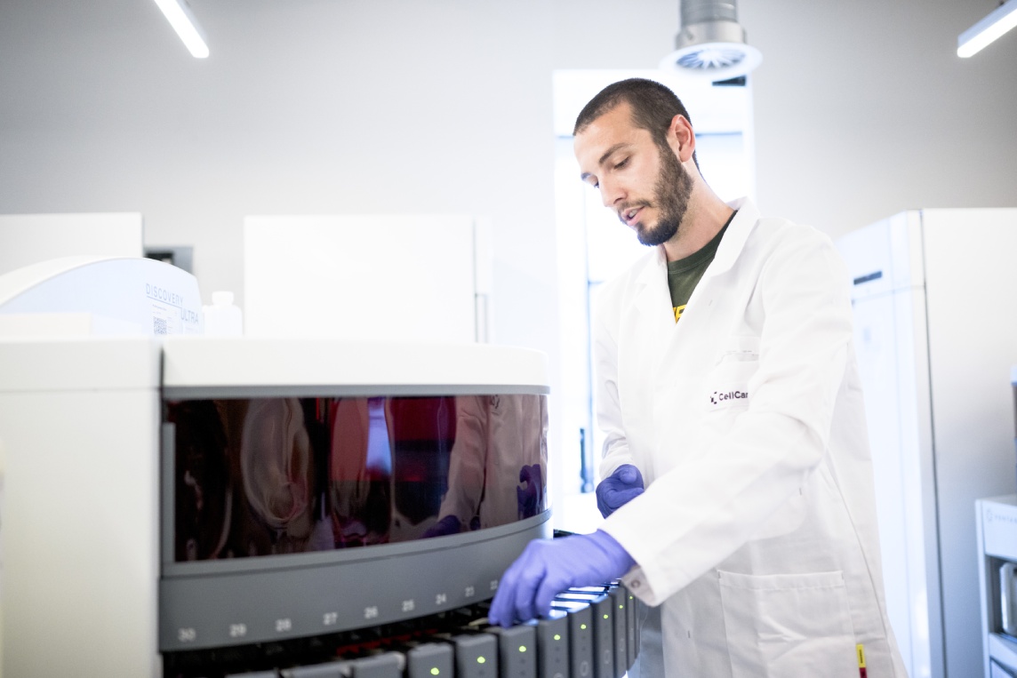
We offer a multitude of brightfield and fluorescent scanners for cell imaging, including:
Our imaging platforms have been implemented in over 1000 clinical trials, and can support your study at the exploratory, primary, and secondary endpoint.
We can perform hematoxylin and eosin (H&E) staining on your clinical samples, allowing you to investigate therapeutic efficacy, including tissue inflammation, tumor-infiltrating lymphocytes (TILs), and tertiary lymphoid structures (TLS).
Our H&E staining services can visualize cellular and tissue structures in FFPE, fresh, and frozen tissue.
All H&E scoring and annotation is performed by our in-house pathologists.
Our pathologists and imaging scientists have expertise implementing AI-supported digital image analysis platforms for dataset evaluation, including:
Our pathologists perform rigorous review and QC of all datasets, ensuring your study’s results are high-quality and reproducible.
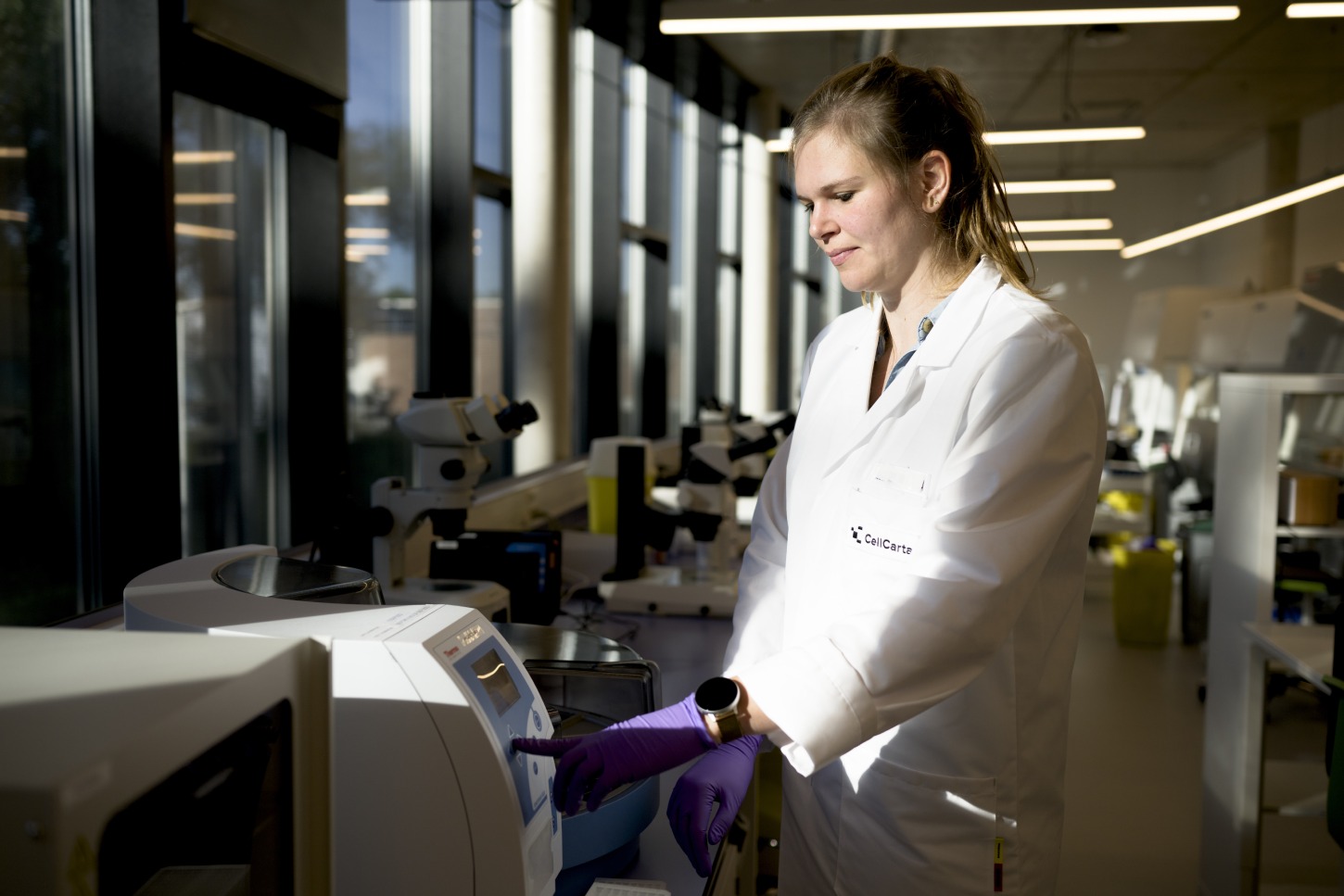
CellCarta’s staining and imaging platforms accommodate a full range of validated IF, IHC, mIF, and mIHC assays, allowing researchers to easily evaluate their biomarkers of choice. Additonally, our team is continually developing new assays and can support any custom solutions your study may require.
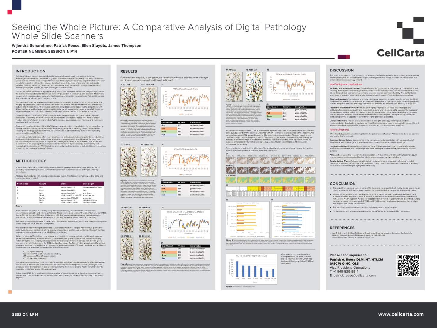
CellCarta evaluated six whole slide scanners across IHC marker panels, quantifying color accuracy and AI algorithm reproducibility across platforms. These findings were verified by our pathologists, demonstrating the rigor needed to select and validate scanners for regulated clinical programs.
Staining and imaging platforms to support histopathology services across all modalities.
Platform Agnostic Approach
Expert In-House Pathologists
Global Execution
AI-Assisted Image Analysis
Cross-Site Harmonization
Staining and imaging platforms built for the specific demands of clinical biomarker programs, from single IHC assays to full digital pathology workflows.
Insights into the abundance, distribution, and localization of your antigens of interest.
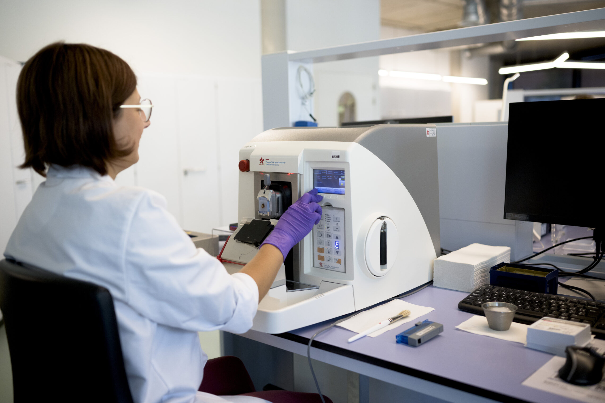
Precise localization and quantification of nucleic acids including DNA, mRNA, and microRNA.
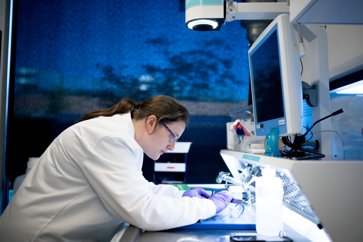
Cutting-edge digital and AI-powered analysis to advance your clinical trials.

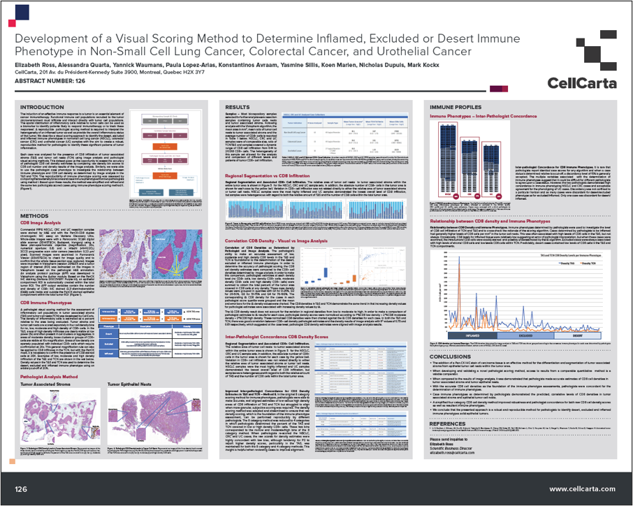 Posters
 Blog
Blog
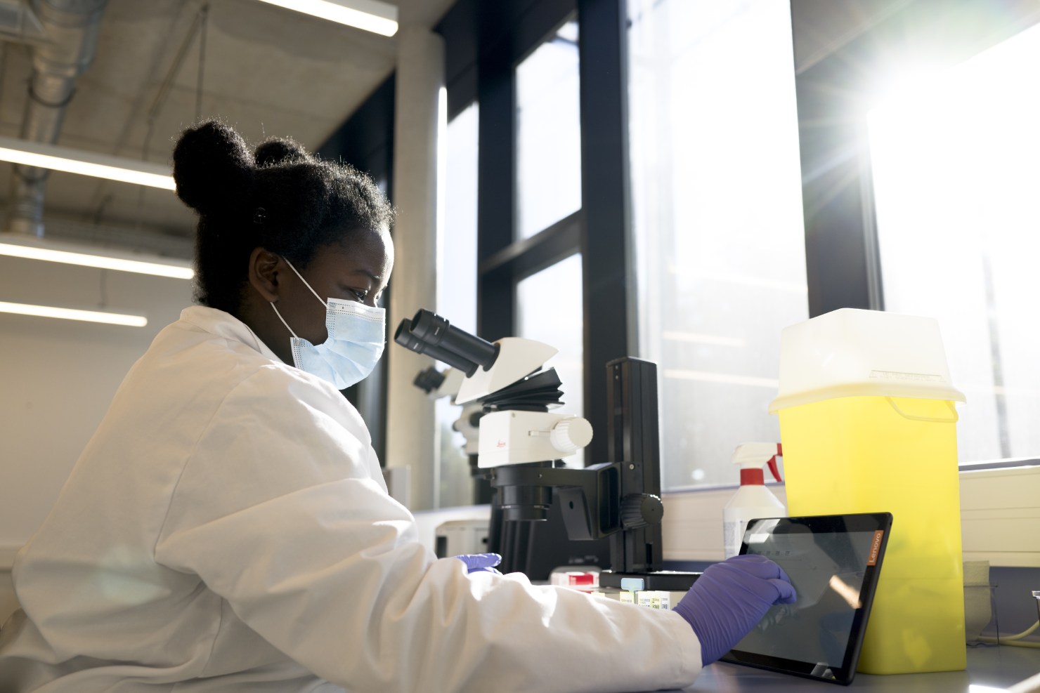
CellCarta runs IHC assays across a multi-instrument staining environment: Dako Autostainer Link 48 and Omnis; Ventana BenchMark Ultra, Ultra Plus, Discovery Ultra, and Special Stains System; Lunaphore COMET; Abbott VIP 2000 and Thermobrite; Sakura Tissue-Tek Prisma; and Leica Biosystems Bond Rx and Bond III.
Chromogenic and fluorescent labeling methods are available across all platforms, giving study teams flexibility to select the instrument format most appropriate for their assay type and tissue. The same instruments and protocols are cross-validated across CellCarta's laboratories in the USA, Belgium, and China, ensuring staining results are directly comparable regardless of which site processes the samples.
CellCarta's imaging infrastructure spans multiple vendors: 3DHistech (Pannoramic 250 and Pannoramic 1000), Ventana (DP 200 and DP 600), Leica Biosystems (GT450 Dx, AT2, and AT Turbo), Lunaphore COMET, RareCyte CyteFinder HT, Zeiss Axio Scan.Z1, Akoya PhenoImager, and BioView (Duet-3 and Allegro Plus). Both brightfield and fluorescent modalities are covered, supporting all standard staining types including IHC, mIF, and ISH. Whole slide scanning is available as a stand-alone digitization service or integrated within a full digital pathology program.
Image analysis is performed using a portfolio of commercially validated AI-assisted software tools: Indica Labs HALO®, Visiopharm®, Lunaphore HORIZON, Akoya inForm®, CyteHub®, SoloWeb, navify® by Roche, and the Lunit SCOPE® suite. These platforms support cell phenotyping, nuclear segmentation, tissue classification, density quantification, and spatial biomarker data extraction. Custom algorithms can be developed for specific clinical programs when commercially available tools do not meet study requirements. All image analysis workflows are 21 CFR Part 11-compliant, supporting data integrity requirements for regulated clinical submissions. An in-house team of 20+ pathologists performs rigorous QC review across all datasets before results are delivered.