November 13, 2025
What is spatial biology?
Understanding how cells interact in their native context is critical for the development of novel therapeutics and effective diagnostics. Conventional assays or single-cell approaches often miss this layer of information, either by averaging signals across the tissue or by removing cells from their tissue environment. Spatial biology, on the other hand, preserves tissue architecture, allowing researchers to study cell type, location, and interaction together to reveal a more complete picture of disease biology.
The value of spatial biology
Spatial biology provides researchers with insights that can directly inform therapeutic development and translational decision-making. By combining cellular detail with spatial context, it enables teams to:
- Map the tissue microenvironment to understand how e.g. immune, stromal, and cancer cells shape disease progression
- Identify new therapeutic targets and validate them in situ, rather than in dissociated systems
- Pinpoint mechanisms of treatment response or resistance by showing how spatial relationships can influence therapeutic effects, giving insight into why patients with seemingly similar profiles can respond differently
- Strengthen biomarker discovery and validation by identifying spatial patterns that correlate with clinical outcomes
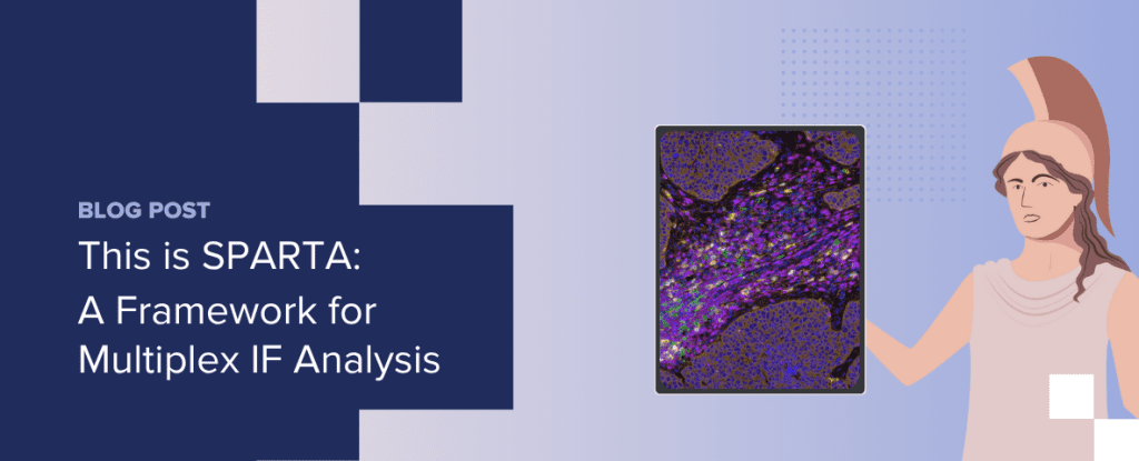
Spatial biology methods generate exceptionally rich datasets that capture multiple layers of tissue complexity. One of the more common spatial biology approaches is multiplex immunofluorescence (mIF), which enables multiple markers to be visualized on a single tissue section. While these datasets bring enormous value, their size and complexity can make them difficult to interpret. When multiple imaging platforms are in use, workflows can become fragmented, and researchers risk generating data that cannot easily be integrated, slowing discovery and translation.
Realizing the full value of spatial biology requires standardized multiplex immunofluorescence protocols and structured workflows that can handle complex datasets and deliver clear, interpretable insights.
SPARTA: Bringing order to spatial biology
To simplify mIF workflows, CellCarta developed SPARTA (Spatial Phenotyping and Analysis in Regions of Tissue with AI)—a platform-agnostic framework that standardizes how datasets are processed and analyzed across imaging systems.
SPARTA incorporates:
- Whole-slide imaging and QC across multiple platforms (Lunaphore Comet, Akoya PhenoImager, and Zeiss Axioscan Z1), offering flexibility and shorter lead times
- Segmentation and classification, using expert-designed, AI-enabled algorithms for segmentation, followed by single-cell feature extraction and rule-based classification
- Data export and advanced analytics, including summary outputs (densities, proportions), object-based single-cell data with coordinates, and advanced analyses such as supervised/unsupervised phenotyping, proximity analysis, and cell neighborhood mapping
- Custom data delivery that avoids generic “data dumps,” providing instead tailored insights aligned with specific research questions
By combining platform flexibility, advanced analytics, and tailored reporting, the SPARTA framework ensures that data from different platforms is processed consistently, so researchers can more easily extract meaningful patterns from complex datasets.
Drive discovery with spatial biology
By revealing the organization of complex tissue environments and capturing cellular interactions, spatial biology continues to provide critical insights that shape drug development. mIF makes it possible to generate rich, multi-dimensional datasets that inform preclinical research and translational decisions. With structured workflows like SPARTA, supported by strong expertise and collaborations across assay development, pathology, imaging, and data science teams, this data can be transformed into tailored outputs that can help accelerate discovery, strengthen biomarker programs, and support clinical development.
About the author

Yannick Waumans is Executive Director of Histopathology Operations at CellCarta, where he oversees the Histopathology Lab Services, Technical Transfer Office, and Digital Pathology Solutions. With a PhD in Pharmaceutical Sciences and over a decade of experience in histopathology, imaging, and image analysis, Yannick is an expert in advancing and operationalizing digital pathology technologies.
He is especially passionate about leveraging digital pathology to accelerate research and improve clinical outcomes.
You might also be interested by
Press Releases
CellCarta and Pillar Biosciences Announce Multi-Year Global Strategic Partnership to Expand Rapid, Decentralized Tumor Profiling for Oncology Clinical Trials
April 7, 2026
More infoNews
CellCarta and Lunit Announce Strategic Collaboration to Accelerate AI-Enabled Digital Pathology for Companion Diagnostic Programs
March 30, 2026
More info